Search Thermo Fisher Scientific
Disclaimer

Clicking the images or links will redirect you to a website hosted by BenchSci that provides third-party scientific content. Neither the content nor the BenchSci technology and processes for selection have been evaluated by us; we are providing them as-is and without warranty of any kind, including for use or application of the Thermo Fisher Scientific products presented.
Invitrogen
CD8a Monoclonal Antibody (OX8), Super Bright™ 780, eBioscience™
")
FIGURE: 1 / 1
CD8a Antibody (78-0084-80) in Flow
Wistar rat splenocytes were stained with CD4 Monoclonal Antibody, FITC (Product # 11-0040-82) and 1.0 µg of Mouse IgG1 kappa Isotype Control, Super Bright 780 (Product # 78-4714-82) (left) or 1.0 µg of CD8a Monoclonal Antibody, Super Bright 780 (right). Cells in the lymphocyte gate were used for analysis.

 in Flow")
Product Details
78-0084-80
Product Specifications
Species Reactivity
Rat
Host/Isotype
Mouse
/ IgG1, kappa
Recommended Isotype Control
Class
Monoclonal
Type
Antibody
Clone
OX8
Conjugate
Excitation/Emission Max
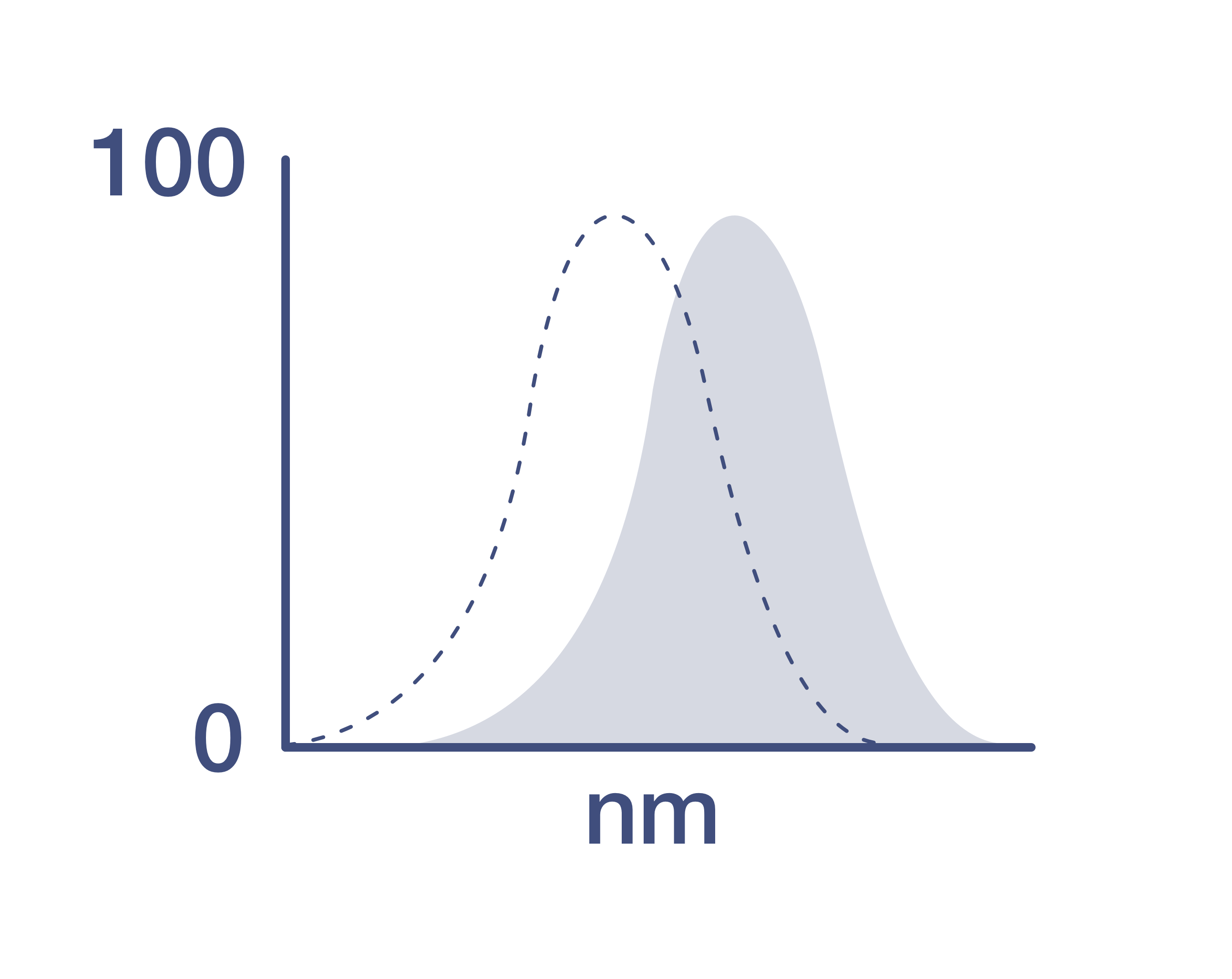
413/780 nm
View spectra
Form
Liquid
Concentration
0.2 mg/mL
Purification
Affinity chromatography
Storage buffer
PBS, pH 7.2, with BSA
Contains
0.09% sodium azide
Storage conditions
4° C, store in dark, DO NOT FREEZE!
Shipping conditions
Ambient (domestic); Wet ice (international)
RRID
AB_2784895
Product Specific Information
Description: The OX8 monoclonal antibody reacts with rat CD8 alpha. CD8 alpha is a member of the immunoglobulin superfamily which heterodimerizes with CD8 beta to form the CD8 antigen expressed on CD4+CD8+, double-positive thymocytes, the majority of MHC class I-restricted peripheral T cells, and on some macrophages. CD8 binds to MHC class I expressed on the surface of antigen-presenting cells during antigen presentation, and participates in T-cell receptor signal transduction through association with the kinase Lck.
Applications Reported: This OX8 antibody has been reported for use in flow cytometric analysis.
Applications Tested: This OX8 antibody has been tested by flow cytometric analysis of rat splenocytes. This may be used at less than or equal to 1.0 µg per test. A test is defined as the amount (µg) of antibody that will stain a cell sample in a final volume of 100 µL. Cell number should be determined empirically but can range from 10^5 to 10^8 cells/test. It is recommended that the antibody be carefully titrated for optimal performance in the assay of interest.
Super Bright 780 is a tandem dye that can be excited with the violet laser line (405 nm) and emits at 780 nm. We recommend using a 780/60 bandpass filter. Please make sure that your instrument is capable of detecting this fluorochrome.
In some experiments, we have observed that compensation values for Super Bright 780-conjugated antibodies are higher in the violet 450/50 channel when using UltraComp eBeads microspheres (Product # 01-2222-42) as compared to single-color stained cells. In such circumstances, we would recommend setting compensation with cells. We have also observed this in some experiments using AbC Total Antibody Compensation beads (Product # A10497).
When using two or more Super Bright dye-conjugated antibodies in a staining panel, it is recommended to use Super Bright Complete Staining Buffer (Product # SB-4401) to minimize any non-specific polymer interactions. Please refer to the datasheet for Super Bright Staining Buffer for more information.
Light sensitivity: This tandem dye is sensitive to photo-induced oxidation. Please protect this vial and stained samples from light.
Fixation: Samples can be stored in IC Fixation Buffer (Product # 00-8222-49) (100 µL of cell sample + 100 µL of IC Fixation Buffer) or 1-step Fix/Lyse Solution (Product # 00-5333-57) for up to 3 days in the dark at 4°C with minimal impact on brightness and FRET efficiency/compensation. Some generalizations regarding fluorophore performance after fixation can be made, but clone specific performance should be determined empirically.
Excitation: 405 nm; Emission: 780 nm; Laser: Violet Laser
Super Bright Polymer Dyes are sold under license from Becton, Dickinson and Company.
Target Information
Cluster of differentiation 8 (CD8), a type I transmembrane glycoprotein of the immunoglobulin family of receptors, plays an integral role in signal transduction, and T cell differentiation and activation. CD8 is predominantly expressed on T cells as a disulfide-linked heterodimer of CD8alpha and CD8beta, where it functions as a co-receptor, along with T cell receptor (TCR), for major histocompatibilty complex class I (MHC-I) molecules; whereas its counterpart, CD4, acts as a co-receptor for MHC-II molecules. CD8 exists on the cell surface, where the CD8alpha chain is essential for binding to MHC-I. CD8 is also expressed on a subset of T cells, NK cells, monocytes and dendritic cells as disulfide-linked homodimers of CD8alpha. Ligation of MHC-I/peptide complexes presented by antigen-presenting cells (APCs), triggers the recruitment of lymphocyte-specific protein tyrosine kinase (Lck), which leads to lymphokine production, motility and cytotoxic T lymphocyte (CTL) activation. Once activated, CTLs play a crucial role in the clearance of pathogens and tumor cells. Differentiation of naive CD8+ T cells into CTLs is strongly enhanced by IL-2, IL-12 and TGF-beta1.
For Research Use Only. Not for use in diagnostic procedures. Not for resale without express authorization.
How to use the Panel Builder
Watch the video to learn how to use the Invitrogen Flow Cytometry Panel Builder to build your next flow cytometry panel in 5 easy steps.
References (0)
Have you cited this product in a publication?
Let us know so we can reference it here.
Bioinformatics
Protein Aliases: CD8 antigen 32 kDa chain; CD8 antigen 37 kDa chain; CD8 antigen alpha-chain; CD8 antigen beta-chain; CD8 antigen, alpha chain; CD8 antigen, alpha-chain; CD8 antigen, beta chain; CD8 antigen, beta-chain; CD8a; CD8alpha; CD8b; CD8beta; fCD8; Leu-2; leu-2a; OX-8 membrane antigen; T-cell surface glycoprotein CD8 alpha chain; T-cell surface glycoprotein CD8 beta chain
Gene Aliases: Cd8a; Cd8b; Cd8b1
UniProt ID: (Rat) P07725, (Rat) P05541
Entrez Gene ID: (Rat) 24930, (Rat) 24931

Performance Guarantee
If an Invitrogen™ antibody doesn't perform as described on our website or datasheet,we'll replace the product at no cost to you, or provide you with a credit for a future purchase.*
Learn more
We're here to help
Get expert recommendations for common problems or connect directly with an on staff expert for technical assistance related to applications, equipment and general product use.
Contact tech support