Search Thermo Fisher Scientific
Disclaimer

Clicking the images or links will redirect you to a website hosted by BenchSci that provides third-party scientific content. Neither the content nor the BenchSci technology and processes for selection have been evaluated by us; we are providing them as-is and without warranty of any kind, including for use or application of the Thermo Fisher Scientific products presented.
Invitrogen
CD274 (PD-L1, B7-H1) Monoclonal Antibody (MIH5), NovaFluor™ Red 700, eBioscience™
 Antibody in Flow Cytometry (Flow)")
FIGURE: 1 / 2
CD274 (PD-L1, B7-H1) Antibody (M036T02R03) in Flow
C57BL/6 mouse splenocytes were unstained (left) or stained with 0.4 µg of CD274 (PD-L1, B7-H1) Monoclonal Antibody, NovaFluor Red 700 (Product # M036T03R03) (right). All cells were co-stained with CD3e Monoclonal Antibody, eFluor 450 (Product # 48-0031-82). Total viable cells in the lymphocyte gate were used for analysis, as determined by LIVE/DEAD Blue (Product # L34962). Data was acquired on a 5-laser Cyt... View More


 Antibody (M036T02R03) in Flow")
 Antibody (M036T02R03) in Flow")
Product Details
M036T02R03
Product Specifications
Host/Isotype
Rat
/ IgG2a, lambda
Class
Monoclonal
Type
Antibody
Clone
MIH5
Conjugate
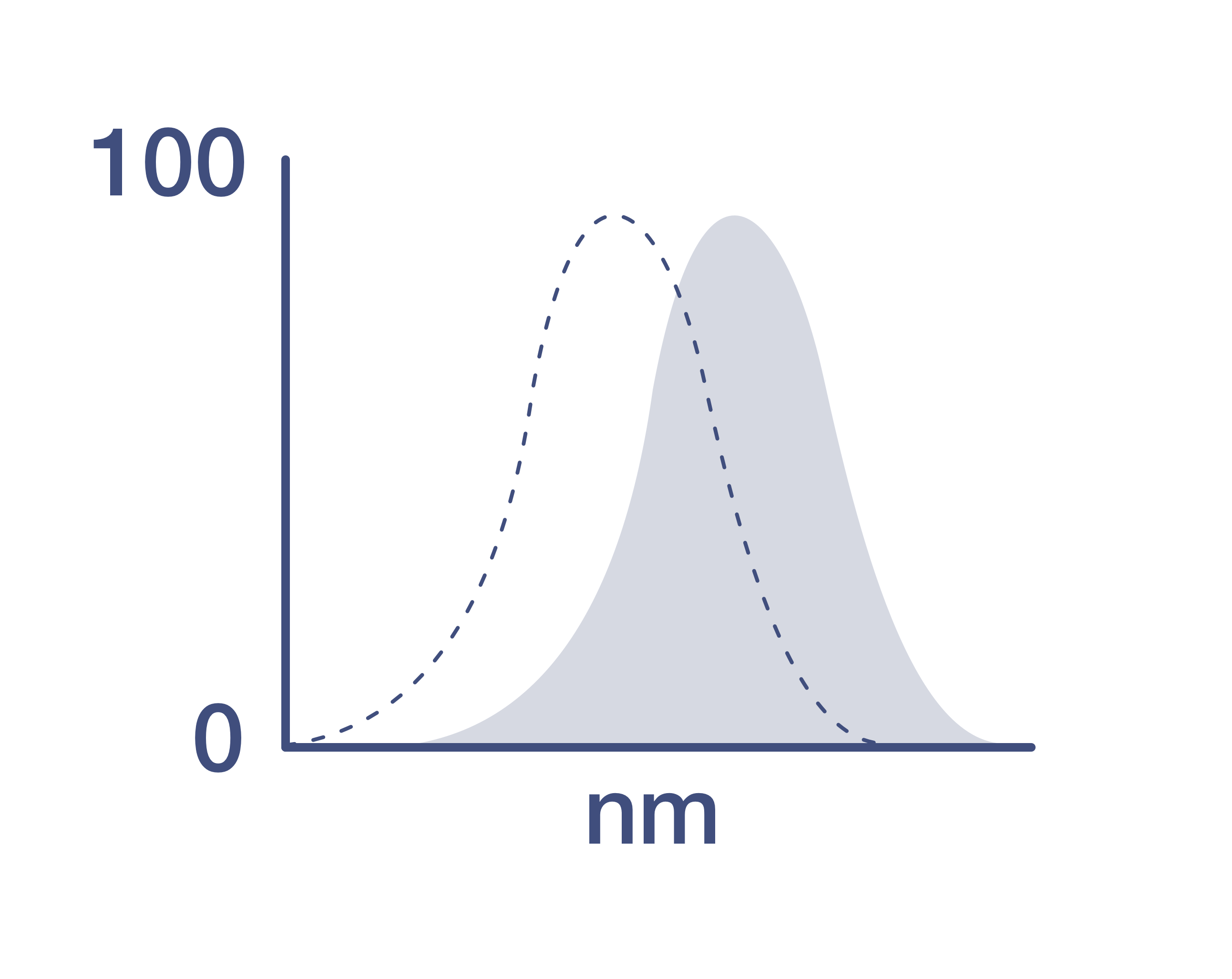
Excitation/Emission Max
637/701 nm
View spectra
Form
Liquid
Concentration
0.1 mg/mL
Storage conditions
4° C, store in dark, DO NOT FREEZE!
Shipping conditions
Ambient (domestic); Wet ice (international)
RRID
AB_2926231
Product Specific Information
Description: The MIH5 monoclonal antibody reacts with mouse B7-H1, also known as PD-L1. B7-H1, a member of the B7 family, has a predicted molecular weight of approximately 40 kDa and belongs to the Ig superfamily. B7-H1 is expressed on a majority of leukocytes including T, B, NK and DC. B7-H1 is a ligand for PD-1. Interaction of PD-1 with either PD-L1 (B7-H1) or PD-L2 (B7-DC) results in inhibition of T and B cell responses. MIH5 is reported to be a blocking antibody.
Applications Reported: The MIH5 antibody has been reported for use in flow cytometric analysis.
Applications Tested: The MIH5 antibody has been tested by flow cytometric analysis of mouse splenocytes. This can be used at less than or equal to 0.2 µg per test. A test is defined as the amount (µg) of antibody that will stain a cell sample in a final volume of 100 µL. Cell number should be determined empirically but can range from 10^5 to 10^8 cells/test. It is recommended that the antibody be carefully titrated for optimal performance in the assay of interest.
NovaFluor dyes are not compatible with DNA intercalating viability dyes. Do not use viability dyes such as propidium iodide, 7-actinomycin D (7-AAD) and DAPI. Invitrogen LIVE/DEAD Fixable Dead Cell stains are recommended for use with NovaFluor dyes.
Each NovaFluor conjugate or kit is shipped with CellBlox Blocking Buffer. Use this buffer whenever staining with NovaFluor conjugates, including single-color compensation controls using cells. Whenever possible, we recommend adding CellBlox Blocking Buffer to antibody cocktails/master mixes prior to combining with cells. Add 5 µL per sample (regardless of the number of NovaFluors in your panel) to use the antibody cocktail as intended. For single-color controls, use 5 µL of CellBlox Blocking Buffer per 100µL of cell sample containing 10^3 to 10^8 cells.
NovaFluor conjugates are based on Phiton™ technology utilizing novel nucleic acid dye structures that allow for engineered fluorescent signatures with consideration for spillover and spread impacts. Learn more
Excitation: 639 nm; Emission: 700 nm; Laser: 633-640 nm (Red) Laser
Target Information
Programmed death receptor ligand 1 (PD-L1, also called B7-H1) is a recently described B7 family member. To date, one specific receptor has been identified that can be ligated by PD-L1. This receptor, programmed death receptor 1 (PD-1), has been shown to negatively regulate T-cell receptor (TCR) signaling. Upon ligating its receptor, PD-L1 has been reported to decrease TCR-mediated proliferation and cytokine production. PD-L1 expression was found to be abundant on many murine and human cancers and could be further up-regulated upon IFN-gamma stimulation. Thus, PD-L1 might play an important role in tumor immune evasion.
For Research Use Only. Not for use in diagnostic procedures. Not for resale without express authorization.
How to use the Panel Builder
Watch the video to learn how to use the Invitrogen Flow Cytometry Panel Builder to build your next flow cytometry panel in 5 easy steps.
References (0)
Have you cited this product in a publication?
Let us know so we can reference it here.

Performance Guarantee
If an Invitrogen™ antibody doesn't perform as described on our website or datasheet,we'll replace the product at no cost to you, or provide you with a credit for a future purchase.*
Learn more
We're here to help
Get expert recommendations for common problems or connect directly with an on staff expert for technical assistance related to applications, equipment and general product use.
Contact tech support