Search Thermo Fisher Scientific
Disclaimer

Clicking the images or links will redirect you to a website hosted by BenchSci that provides third-party scientific content. Neither the content nor the BenchSci technology and processes for selection have been evaluated by us; we are providing them as-is and without warranty of any kind, including for use or application of the Thermo Fisher Scientific products presented.
Invitrogen
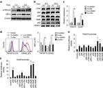
CD252 (OX40 Ligand) Monoclonal Antibody (RM134L), PerCP-eFluor™ 710, eBioscience™
 Antibody in Flow Cytometry (Flow)")
FIGURE: 1 / 7
CD252 (OX40 Ligand) Antibody (46-5905-82) in Flow
Mouse splenocytes were stimulated for 4 days with F (ab')2 Anti-Mouse IgM, u chain specific and Anti-Mouse CD40 Functional Grade Purifieds (Product # 16-5092-85 and Product # 16-0401-82). Cells were stained with 0.125 µg of Rat IgG2b K Isotype Control PerCP-eFluor® 710 (Product # 46-4031-82) (blue histogram) or 0.125 µg of Anti-Mouse CD252 (OX40 Ligand) PerCP-eFluor® 710 (purple histogram). Total viable cel... View More


 Antibody (46-5905-82) in Flow")
 Antibody (46-5905-82) in Flow")
 Antibody (46-5905-82) in Flow")
 Antibody (46-5905-82) in Flow")
 Antibody (46-5905-82) in Flow")
 Antibody (46-5905-82) in Flow")
 Antibody (46-5905-82) in Flow")
Product Details
46-5905-82
Applications
Tested Dilution
Publications
Product Specifications
Species Reactivity
Mouse
Published species
Human
Host/Isotype
Rat
/ IgG2b, kappa
Recommended Isotype Control
Class
Monoclonal
Type
Antibody
Clone
RM134L
Conjugate
PerCP-eFluor™ 710
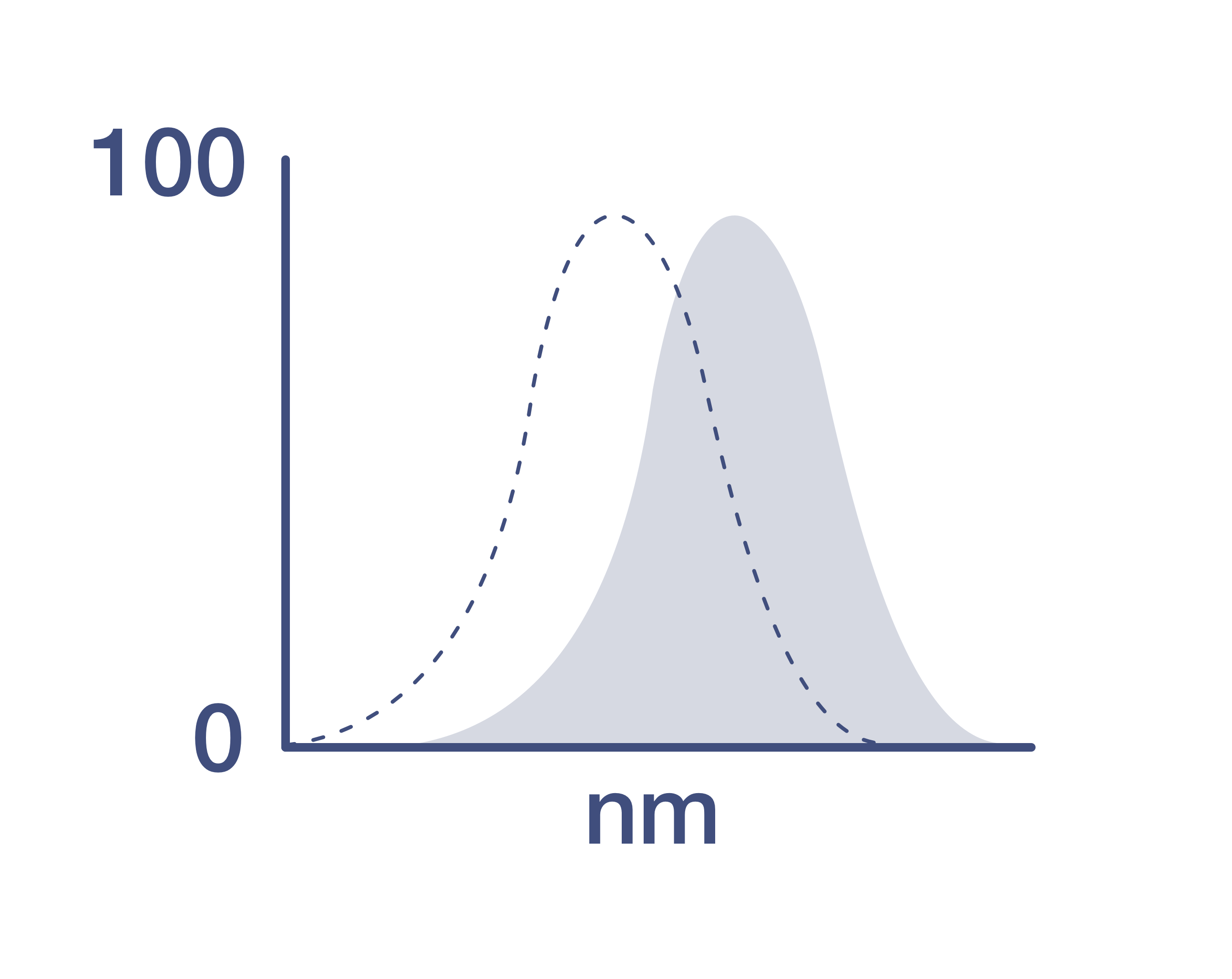
Excitation/Emission Max
482/708 nm
View spectra
Form
Liquid
Concentration
0.2 mg/mL
Purification
Affinity chromatography
Storage buffer
PBS, pH 7.2
Contains
0.09% sodium azide
Storage conditions
4° C, store in dark, DO NOT FREEZE!
Shipping conditions
Ambient (domestic); Wet ice (international)
RRID
AB_2573809
Product Specific Information
Description: The RM134L monoclonal antibody reacts with mouse CD252 also known as OX-40 Ligand, a member of the TNF superfamily. OX-40L is induced on mouse splenic B cells stimulated with a combination of anti-IgM and anti-CD40. Neither resting nor activated mouse T cells express OX-40L. The interaction of OX-40 (CD134) with OX-40L has been implicated in T cell-dependent humoral response. RM134L inhibits the costimulatory activity of mouse OX-40L transfected cells for anti-CD3-stimulated T cell proliferation.
Applications Reported: This RM134L antibody has been reported for use in flow cytometric analysis.
Applications Tested: This RM134L antibody has been tested by flow cytometric analysis of stimulated mouse splenocytes. This can be used at less than or equal to 0.25 µg per test. A test is defined as the amount (µg) of antibody that will stain a cell sample in a final volume of 100 µL. Cell number should be determined empirically but can range from 10^5 to 10^8 cells/test. It is recommended that the antibody be carefully titrated for optimal performance in the assay of interest.
PerCP-eFluor® 710 emits at 710 nm and is excited with the blue laser (488 nm); it can be used in place of PerCP-Cyanine5.5. We recommend using a 710/50 bandpass filter, however, the 695/40 bandpass filter is an acceptable alternative. Please make sure that your instrument is capable of detecting this fluorochrome.
Light sensitivity: This tandem dye is sensitive to photo-induced oxidation. Please protect this vial and stained samples from light.
Fixation: Samples can be stored in IC Fixation Buffer (Product # 00-8222) (100 µL of cell sample + 100 µL of IC Fixation Buffer) or 1-step Fix/Lyse Solution (Product # 00-5333) for up to 3 days in the dark at 4°C with minimal impact on brightness and FRET efficiency/compensation. Some generalizations regarding fluorophore performance after fixation can be made, but clone specific performance should be determined empirically.
Excitation: 488 nm; Emission: 710 nm; Laser: Blue Laser.
Filtration: 0.2 µm post-manufacturing filtered.
Target Information
The tumor necrosis factor superfamily member TNFSF4 is a type II membrane bound, non-covalently linked homotrimeric protein. It is expressed on antigen presenting cells, such as dendritic cells and activated B-cells, and also on other cells such as vascular endothelial cells, mast cells, and natural killer cells. TNFSF4 signals specifically through the TNFRSF4 receptor, is expressed predominantly on CD4+T cells but also on certain activated CD8+T cells. TNFRSF4/TNFSF4 functions as a costimulatory signal, which is required for a productive interaction between antigen presenting cells and their target T-cells. It enhances cell proliferation and survival, and increases expression of RANTES, IL-2, IL-3, and IFNgamma. TNFRSF4/TNFSF4 signaling plays an important role in immunotolerance.
For Research Use Only. Not for use in diagnostic procedures. Not for resale without express authorization.
Bioinformatics
Protein Aliases: atherosclerosis 1; CD252; OX40 ligand; OX40L; tax-transcriptionally activated glycoprotein 1 ligand; Tumor necrosis factor ligand superfamily member 4
Gene Aliases: Ath-1; Ath1; CD134L; gp34; OX-40L; Ox40l; Tnfsf4; TXGP1; Txgp1l
UniProt ID: (Mouse) P43488
Entrez Gene ID: (Mouse) 22164
blood vessel development
cytokine production
defense response to nematode
acute inflammatory response
positive regulation of immune effector process
positive regulation of T cell cytokine production
regulation of adaptive immune response
positive regulation of type 2 immune response
positive regulation of immunoglobulin mediated immune response
inflammatory response
immune response
cholesterol metabolic process
response to virus
negative regulation of interferon-gamma production
negative regulation of interleukin-17 production
positive regulation of interferon-gamma production
positive regulation of interleukin-10 production
positive regulation of interleukin-12 production
positive regulation of interleukin-13 production
positive regulation of interleukin-2 production
positive regulation of interleukin-4 production
positive regulation of interleukin-6 production
memory T cell activation
T-helper 2 cell activation
response to nitrogen dioxide
cellular response to nitrogen dioxide
CD4-positive, alpha-beta T cell costimulation
T cell proliferation
positive regulation of activated T cell proliferation
positive regulation of CD4-positive, alpha-beta T cell differentiation
positive regulation of memory T cell differentiation
negative regulation of sequence-specific DNA binding transcription factor activity
innate immune response
negative regulation of regulatory T cell differentiation
negative regulation of T-helper 1 cell differentiation
positive regulation of T-helper 2 cell differentiation
negative regulation of transcription, DNA-templated
positive regulation of alpha-beta T cell proliferation
negative regulation of cytokine secretion
regulation of inflammatory response
positive regulation of inflammatory response
positive regulation of B cell activation
positive regulation of immunoglobulin secretion
negative regulation of T cell apoptotic process
negative regulation of activation-induced cell death of T cells
cellular response to lipopolysaccharide
cellular response to prostaglandin E stimulus
chemokine (C-C motif) ligand 11 production
positive regulation of CD4-positive, alpha-beta T cell costimulation
positive regulation of T cell migration
positive regulation of T cell costimulation
positive regulation of memory T cell activation
positive regulation of T-helper 2 cell activation
positive regulation of interleukin-4-dependent isotype switching to IgE isotypes
negative regulation of extrinsic apoptotic signaling pathway

Performance Guarantee
If an Invitrogen™ antibody doesn't perform as described on our website or datasheet,we'll replace the product at no cost to you, or provide you with a credit for a future purchase.*
Learn more
We're here to help
Get expert recommendations for common problems or connect directly with an on staff expert for technical assistance related to applications, equipment and general product use.
Contact tech support