Search Thermo Fisher Scientific
Disclaimer

Clicking the images or links will redirect you to a website hosted by BenchSci that provides third-party scientific content. Neither the content nor the BenchSci technology and processes for selection have been evaluated by us; we are providing them as-is and without warranty of any kind, including for use or application of the Thermo Fisher Scientific products presented.
Invitrogen
CD8a Monoclonal Antibody (OKT8 (OKT-8)), NovaFluor™ Blue 800, eBioscience™
Application
FIGURE: 1 / 2
CD8a Antibody (H003T03B12-A) in Flow
Normal human peripheral blood cells were unstained (left) or stained with CD8a Monoclonal Antibody, NovaFluor Blue 800 (right). All cells were co-stained with CD19 Monoclonal Antibody, eFluor 450 (Product # 48-0199-42). Total viable cells in the lymphocyte gate were used for analysis, as determined by LIVE/DEAD Blue (Product # L34962). Data was acquired on a 5-laser Cytek Aurora and unmixed with multiple autofluorescence signature extraction.


 in Flow")
 in Flow")
Product Details
H003T03B12-A
Product Specifications
Species Reactivity
Human
Host/Isotype
Mouse
/ IgG2a, kappa
Class
Monoclonal
Type
Antibody
Clone
OKT8 (OKT-8)
Conjugate
Excitation/Emission Max
493/806 nm
View spectra
Form
Liquid
Concentration
0.1 µg/Test
Purification
Affinity chromatography
Storage buffer
PBS, pH 7.2, with BSA
Contains
0.09% sodium azide
Storage conditions
4°C, store in dark, DO NOT FREEZE!
RRID
AB_3679269
Product Specific Information
Description
The OKT8 monoclonal antibody reacts with the human CD8a molecule, an approximately 32-34 kDa cell surface receptor expressed either as a heterodimer with the CD8 beta chain (CD8 alpha beta) or as a homodimer (CD8 alphaalpha). A majority of thymocytes and a subpopulation of mature T cells and NK cells express CD8a. CD8 binds to MHC class I and through its association with protein tyrosine kinase p56lck plays a role in T-cell development and activation of mature T cells. Preliminary testing indicates that OKT8 and two other mouse anti-human CD8 antibodies (clone RPA-T8, Cat. No.14-0088 and clone HIT8a, Cat. No.14-0089) do not compete with each other for binding to human peripheral blood leukocytes by flow cytometric analysis, suggesting that they do not bind to similar epitopes or block each other by steric hindrance.
This product contains 1 vial of NovaFluor conjugate and 1 vial of CellBlox Plus Blocking Buffer.
Applications Tested
This OKT8 (OKT-8) antibody has been pre-titrated and tested by flow cytometric analysis of normal human peripheral blood cells. This can be used at 5 µL (0.1 µg) per test. A test is defined as the amount (µg) of antibody that will stain a cell sample in a final volume of 100 µL. Cell number should be determined empirically but can range from 10^5 to 10^8 cells/test.
Master mixes
• Master mixes of NFs should be made at 2-8 °C and may be made up to 4 hours ahead of time.
• We do not recommend storing master mixes containing NovaFluor conjugates overnight or longer.
Whole Blood compatibility
• When utilizing whole blood (as opposed to density-gradient-purified PBMC), we recommend lysing red blood cells in bulk prior to staining with NovaFluor conjugates.
• See the Bulk Lysis of Human Whole Blood protocol here.
• Staining of whole blood with NovaFluor conjugates followed by lysis of red blood cells may result in higher-than-expected background staining.
Viability dye compatibility
• NovaFluor dyes are not compatible with DNA intercalating viability dyes.
• Do not use viability dyes such as propidium iodide, 7-actinomycin D (7-AAD) and DAPI. Invitrogen LIVE/DEAD Fixable Dead Cell stains are recommended for use with NovaFluor dyes.
CellBlox Plus Blocking Buffer
• This NovaFluor conjugate comes with CellBlox Plus Blocking Buffer (Cat. No. C001T03F01), essential for optimal staining.
• Use CellBlox Plus Blocking Buffer in all experiments with NovaFluor conjugates.
• Add 5 μL per sample to antibody cocktails/master mixes (regardless of how many Novafluor-conjugated antibodies are present) before combining with cells.
• CellBlox Plus Blocking Buffer is compatible with either Super Bright Complete Blocking Buffer or Brilliant Stain Buffer and can be used in antibody cocktails/master mixes with those reagents.
• For single-color controls, use 5 μL of CellBlox Plus Blocking Buffer per 100 μL of cell sample (10^3 to 10^8 cells).
NovaFluor conjugates are based on Phiton technology utilizing novel fluorophore-containing nucleic acid dye structures that allow for engineered fluorescent signatures with consideration for spillover and spread impacts. Learn more
Excitation: 493 nm; Emission: 806 nm; Laser: 488 nm (Blue) Laser
Target Information
CD8, also known as cluster of differentiation 8, is a type I transmembrane glycoprotein of the immunoglobulin family that plays a crucial role in T cell differentiation, activation, and signal transduction. It is expressed as either a heterodimer (CD8 alpha beta) or a homodimer (CD8 alpha alpha). The CD8 alpha beta form is predominantly found on the majority of thymocytes and a subpopulation of mature alpha beta TCR T cells, while the CD8 alpha alpha form is expressed on gamma delta TCR T cells, a subset of intestinal intraepithelial lymphocytes (IELs), and dendritic cells. CD8 functions as a co-receptor for major histocompatibility complex class I (MHC-I) molecules, working alongside the T cell receptor (TCR). The CD8 alpha chain is essential for binding to MHC-I. CD8 is also expressed on a subset of T cells, NK cells, monocytes, and dendritic cells as disulfide-linked homodimers of CD8 alpha. Upon ligation of MHC-I/peptide complexes presented by antigen-presenting cells (APCs), CD8 recruits lymphocyte-specific protein tyrosine kinase (Lck), leading to lymphokine production, increased motility, and activation of cytotoxic T lymphocytes (CTLs). Activated CTLs are vital for clearing pathogens and tumor cells. The differentiation of naive CD8+ T cells into CTLs is strongly enhanced by cytokines such as IL-2, IL-12, and TGF-beta1. Through its interactions with MHC-I and association with protein tyrosine kinase p56lck, CD8 plays a significant role in T cell development and the activation of mature T cells.
For Research Use Only. Not for use in diagnostic procedures. Not for resale without express authorization.
Video Player is loading.
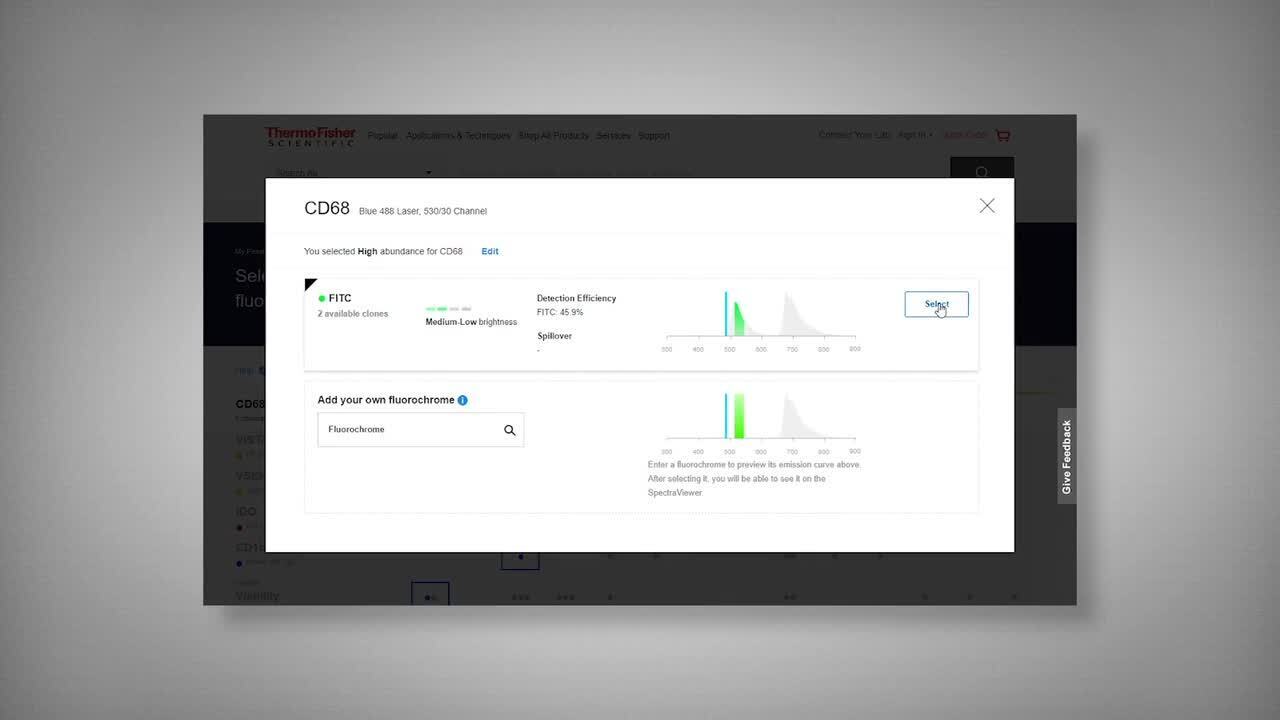
How to use the Panel Builder
Watch the video to learn how to use the Invitrogen Flow Cytometry Panel Builder to build your next flow cytometry panel in 5 easy steps.
References (0)
Have you cited this product in a publication?
Let us know so we can reference it here.
Bioinformatics
Protein Aliases: CD8 antigen, alpha polypeptide (p32); CD8 antigen, beta polypeptide 1 (p37); CD8a; CD8alpha; CD8b; CD8beta; fCD8; Leu-2; leu-2a; Leu2 T-lymphocyte antigen; OKT8 T-cell antigen; T cell co-receptor; T lymphocyte surface glycoprotein beta chain; T-cell antigen Leu2; T-cell surface glycoprotein CD8 alpha chain; T-cell surface glycoprotein CD8 beta chain; T-lymphocyte differentiation antigen T8/Leu-2; T8 T-cell antigen
Gene Aliases: CD8; CD8A; CD8B; CD8B1; LEU2; LY3; LYT3; MAL; p32; P37
UniProt ID: (Human) P01732, (Human) P10966
Entrez Gene ID: (Human) 925, (Human) 926

Performance Guarantee
If an Invitrogen™ antibody doesn't perform as described on our website or datasheet,we'll replace the product at no cost to you, or provide you with a credit for a future purchase.*
Learn more
We're here to help
Get expert recommendations for common problems or connect directly with an on staff expert for technical assistance related to applications, equipment and general product use.
Contact tech support