Search Thermo Fisher Scientific
Disclaimer

Clicking the images or links will redirect you to a website hosted by BenchSci that provides third-party scientific content. Neither the content nor the BenchSci technology and processes for selection have been evaluated by us; we are providing them as-is and without warranty of any kind, including for use or application of the Thermo Fisher Scientific products presented.
Invitrogen
PIR-A/B Monoclonal Antibody (10-1-PIR), APC, eBioscience™
Promotions
View available promotion(s)
Promo Code: P5877545 Don't miss out: buy 3, only pay for 2 Extended to June 20: take advantage of our 3 for 2 promotion. Whether you are restocking essentials or trying something new, now is a great time to get more for your money!. Learn more
Promo Code: RPUZZ25 Stock up on essentials to piece your discovery together Until June 27, save up to $650 and get an exclusive lab-themed hidden-object puzzle. Learn more
Application
FIGURE: 1 / 4
PIR-A/B Antibody (17-3101-82) in Flow
Staining of C57BL/6 splenocytes with Anti-Mouse CD19 FITC (Product # 11-0193-82) and 0.125 µg of Rat IgG2b kappa Isotype Control APC (Product # 17-4031-82) (left) or 0.125 µg of Anti-Mouse PIR-A/B APC (right). Total viable cells were used for analysis.


 in Flow")
 in Flow")
 in Flow")
 in Flow")
Product Details
17-3101-82
Applications
Tested Dilution
Publications
Product Specifications
Species Reactivity
Mouse
Host/Isotype
Rat
/ IgG2b, kappa
Recommended Isotype Control
Class
Monoclonal
Type
Antibody
Clone
10-1-PIR
Conjugate
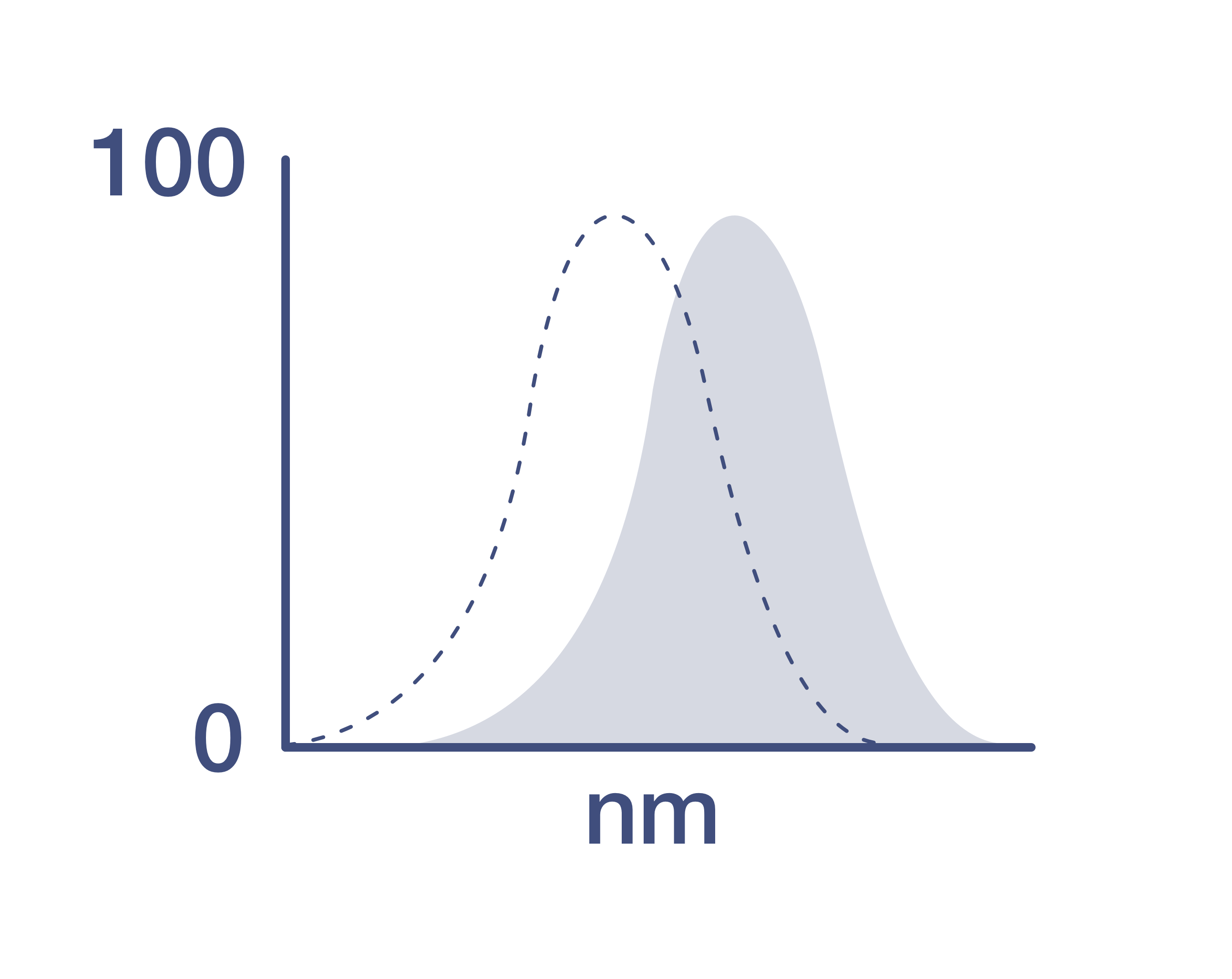
Excitation/Emission Max
651/660 nm
View spectra
Form
Liquid
Concentration
0.2 mg/mL
Purification
Affinity chromatography
Storage buffer
PBS, pH 7.2
Contains
0.09% sodium azide
Storage conditions
4°C, store in dark, DO NOT FREEZE!
Shipping conditions
Ambient (domestic); Wet ice (international)
RRID
AB_1944406
Product Specific Information
Description: This 10-1-PIR monoclonal antibody reacts with mouse paired Ig-like receptors of activating (PIR)-A and -B. These cell surface glycoproteins, which contain six extracellular Ig-like domains with distinct transmembrane and cytoplasmic regions, are expressed together on B lymphocytes, dendritic cells, macrophages, granulocytes, platelets, and mast cells. However, expression of PIR-A/B has not been observed on T cells, NK cells, and erythrocytes. The PIR-A receptor interacts with signaling molecules containing immunoreceptor tyrosine-based activation motifs (ITAMs) which lead to its activating function. In contrast, the PIR-B receptor associates with proteins possessing immunoreceptor tyrosine-based inhibitory motifs (ITIMs); therefore, this receptor has been shown to have an inhibitory function. PIRs bind MHC class I to modulate cell signaling and homeostasis of the immune system. Moreover, PIR-B knockout mice have been shown to exhibit susceptibility to Salmonella infection.
Crossblocking studies indicate that 10-1-PIR recognizes a different epitope from 6C1 (Product # 46-5978).
Applications Reported: This 10-1-PIR antibody has been reported for use in flow cytometric analysis.
Applications Tested: This 10-1-PIR antibody has been tested by flow cytometric analysis of mouse splenocytes. This can be used at less than or equal to 0.25 µg per test. A test is defined as the amount (µg) of antibody that will stain a cell sample in a final volume of 100 µL. Cell number should be determined empirically but can range from 10^5 to 10^8 cells/test. It is recommended that the antibody be carefully titrated for optimal performance in the assay of interest.
Excitation: 633-647 nm; Emission: 660 nm; Laser: Red Laser.
Filtration: 0.2 µm post-manufacturing filtered.
Target Information
Paired Ig-like receptors of activating (PIR)-A and -B are expressed on B lymphocytes, macrophages, dendritic cells, platelets, granulocytes, and mast cells, but not on T cells, NK cells, and erythrocytes. These cell surface glycoproteins contain six extracellular Ig-like domains with distinct cytoplasmic and transmembrane regions. The PIR-A receptor activates through signaling molecules containing immunoreceptor tyrosine-based activation motifs. In contrast, the PIR-B receptor inhibits through proteins possessing immunoreceptor tyrosine-based inhibitory motifs. PIRs bind MHC class I to modulate immune system homeostasis and cell signaling.
For Research Use Only. Not for use in diagnostic procedures. Not for resale without express authorization.
Video Player is loading.
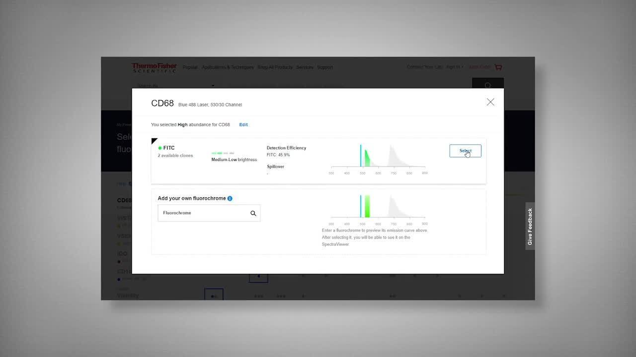
How to use the Panel Builder
Watch the video to learn how to use the Invitrogen Flow Cytometry Panel Builder to build your next flow cytometry panel in 5 easy steps.
Bioinformatics
Protein Aliases: CD85k; Cell-surface glycoprotein p91; Gp49b; leukocyte immunoglobulin-like receptor 3; Leukocyte immunoglobulin-like receptor subfamily B member 3; leukocyte immunoglobulin-like receptor, subfamily B (with TM and ITIM domains), member 3; Lilrb4; LIR-3; paired immunoglobulin-like receptor; Paired immunoglobulin-like receptor B; paired-Ig-like receptor A1; PIR-B
Gene Aliases: 6M21; Gp91; Lilrb3; LIR-3; Ly89; Pir; PIR-A1; PIR-B; Pirb
UniProt ID: (Mouse) P97484
Entrez Gene ID: (Mouse) 18722, (Mouse) 18733

Performance Guarantee
If an Invitrogen™ antibody doesn't perform as described on our website or datasheet,we'll replace the product at no cost to you, or provide you with a credit for a future purchase.*
Learn more
We're here to help
Get expert recommendations for common problems or connect directly with an on staff expert for technical assistance related to applications, equipment and general product use.
Contact tech support