Search Thermo Fisher Scientific
Disclaimer

Clicking the images or links will redirect you to a website hosted by BenchSci that provides third-party scientific content. Neither the content nor the BenchSci technology and processes for selection have been evaluated by us; we are providing them as-is and without warranty of any kind, including for use or application of the Thermo Fisher Scientific products presented.
Invitrogen
phospho-Tyrosine Monoclonal Antibody (pY20), eFluor™ 450, eBioscience™
Application
FIGURE: 1 / 4
phospho-Tyrosine Antibody (48-5001-42) in Flow
Intracellular staining of normal human peripheral blood cells left untreated (orange histogram) or treated with hydrogen-peroxide-activated sodium pervanadate (purple histogram) and stained with 0.06 µg of Anti-Human/Mouse phospho-Tyrosine eFluor® 450 (purple histogram) using the IC Fixation & Permeabilization Buffer Set (Product # 88-8824-00). Cells in the lymphocyte gate were used for analysis.


 in Flow")
 in ICC/IF")
 in ICC/IF")
 in Flow")
Product Details
48-5001-42
Applications
Tested Dilution
Publications
Product Specifications
Species Reactivity
Chemical
Published species
Not Applicable
Host/Isotype
Mouse
/ IgG2b, kappa
Recommended Isotype Control
Class
Monoclonal
Type
Antibody
Clone
pY20
Conjugate
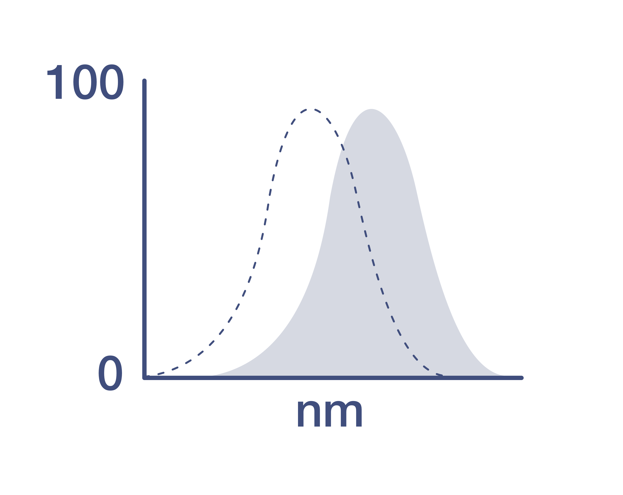
Excitation/Emission Max
405/445 nm
View spectra
Form
Liquid
Concentration
5 µL/Test
Purification
Affinity chromatography
Storage buffer
PBS, pH 7.2, with 0.2% BSA
Contains
0.09% sodium azide
Storage conditions
4° C, store in dark, DO NOT FREEZE!
Shipping conditions
Ambient (domestic); Wet ice (international)
RRID
AB_2574064
Product Specific Information
Description: The pY20 monoclonal antibody recognizes phosphorylated tyrosine residues (p-Tyr) on proteins. Numerous intracellular signaling cascades are propagated via phosphorylation of specific tyrosine on signaling proteins. The detection of p-Tyr residues is valuable for the characterization and purification of phosphorylated proteins and the biochemical pathways that they are involved in.
Applications Reported:This pY20 antibody has been reported for use in intracellular staining followed by flow cytometric analysis.
Applications Tested: This pY20 antibody has been pre-titrated and tested by intracellular staining followed by flow cytometric analysis of normal human peripheral blood cells. This can be used at 5 µL (0.06 µg) per test. A test is defined as the amount (µg) of antibody that will stain a cell sample in a final volume of 100 µL. Cell number should be determined empirically but can range from 10^5 to 10^8 cells/test.
eFluor™ 450 is an alternative to Pacific Blue™. eFluor™ 450 emits at 445 nm and is excited with the Violet laser (405 nm). Please make sure that your instrument is capable of detecting this fluorochrome.
Staining Protocol: All protocols work well for this monoclonal antibody. Use of Protocol A: Two-step protocol: intracellular (cytoplasmic) proteins allows for the greatest flexibility for detection of surface and intracellular (cytoplasmic) proteins. Use of Protocol B: One-step protocol: intracellular (nuclear) proteins is recommended for staining of transcription factors in conjunction with surface and phosphorylated intracellular (cytoplasmic) protein(s). Protocol C: Two-step protocol: Fixation/Methanol allows for the greatest discrimination of phospho-specific signaling between unstimulated and stimulated samples, but with limitations on the ability to stain specific surface proteins (refer to "Clone Performance Following Fixation/Permeabilization" located in the BestProtocols Section under the Resources tab online). All Protocols can be found in the Flow Cytometry Protocols: "Staining Intracellular Antigens for Flow Cytometry Protocol" located in the BestProtocols® Section under the Resources tab online.
Excitation: 405 nm; Emission: 445 nm; Laser: Violet Laser.
Filtration: 0.2 µm post-manufacturing filtered.
Target Information
The role of tyrosine phosphorylation in transduction of the mitogenic signal from transmembrane receptors and in transformation by oncogene tyrosine kinases has been the subject of intense investigation for several years. While the phosphorylation of specific tyrosine residues has been shown to be a primary mechanism of signal transduction during normal mitogenesis, cell cycle progression and oncogenic transformation, its role in other areas such as differentiation and gap junction communication, is a matter of active and ongoing research. Antibodies that specifically recognize phosphorylated tyrosine residues have proved to be invaluable to the study of tyrosine -phosphorylated proteins and the biochemical pathways in which they function. The fluorescein (FITC) conjugate of clone PY20 anti-phosphotyrosine is especially useful for the detection of these P-Tyr proteins in immunohistochemical and immunocytochemical protocols in situations wherein the use of a secondary antibody would complicate detection of the protein(s) of interest.
For Research Use Only. Not for use in diagnostic procedures. Not for resale without express authorization.
Video Player is loading.
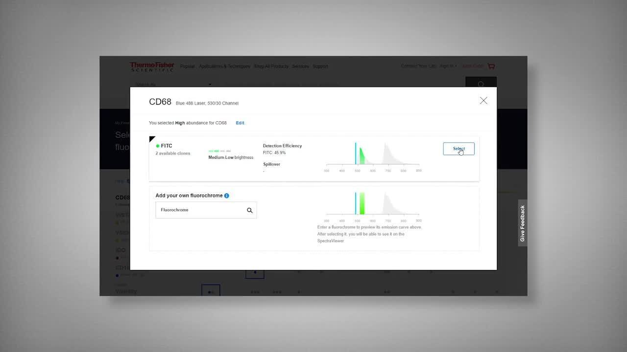
How to use the Panel Builder
Watch the video to learn how to use the Invitrogen Flow Cytometry Panel Builder to build your next flow cytometry panel in 5 easy steps.
Bioinformatics
Protein Aliases: Phosphotyrosine; pTyr; pY

Performance Guarantee
If an Invitrogen™ antibody doesn't perform as described on our website or datasheet,we'll replace the product at no cost to you, or provide you with a credit for a future purchase.*
Learn more
We're here to help
Get expert recommendations for common problems or connect directly with an on staff expert for technical assistance related to applications, equipment and general product use.
Contact tech support