Quattro Environmental Scanning Electron Microscope
The Thermo Scientific Quattro ESEM combines all-around performance in imaging and analytics with a unique environmental mode (ESEM) that allows samples to be studied in their natural state. It is ideal for a wide variety of academic, industrial, and government labs that want the versatility and ease of use needed for multiple users of different experience levels and disciplines on a platform that also supports unique in situ experiments. The Quattro ESEM features a field emission gun (FEG), which ensures excellent resolution. Meanwhile, its three vacuum modes (high vacuum, low vacuum, and ESEM) provide the flexibility to accommodate the widest range of samples of any SEM available, including those that are outgassing or otherwise not vacuum-compatible.
Learn more about this product in our on-demand webinar
Discover how to analyze liquids and hydrated samples in 3D using environmental scanning electron microscopy.
Best in class for in situ imaging capabilities, dynamic experiments with excellent analytical capabilities
The Quattro ESEM is a highly flexible platform for in situ dynamic experiments. The Quattro ESEM's environmental SEM capability allows scientists to study materials in a range of conditions, such as wet/humid, hot, or reactive environments, as they develop new materials and products. It supports cooling and heating experiments both in high vacuum and low vacuum to accommodate the widest range of needs. Cooling experiments on wet materials are possible with the Peltier cooling stage or if the transmission mode is needed, with Thermo Scientific WetSTEM Technology. Additionally, the Quattro ESEM provides several different possibilities to heat the samples of interest, both bulk and powders. Users can perform dynamic experiments at high temperatures both in high vacuum (with the high-vacuum heating stage), in low-vacuum/ESEM mode (with the two ESEM heating stages for temperatures up to 1,000°C and 1,400°C), and on a localized area for better control of the temperature change (with the Thermo Scientific µHeater Holder).
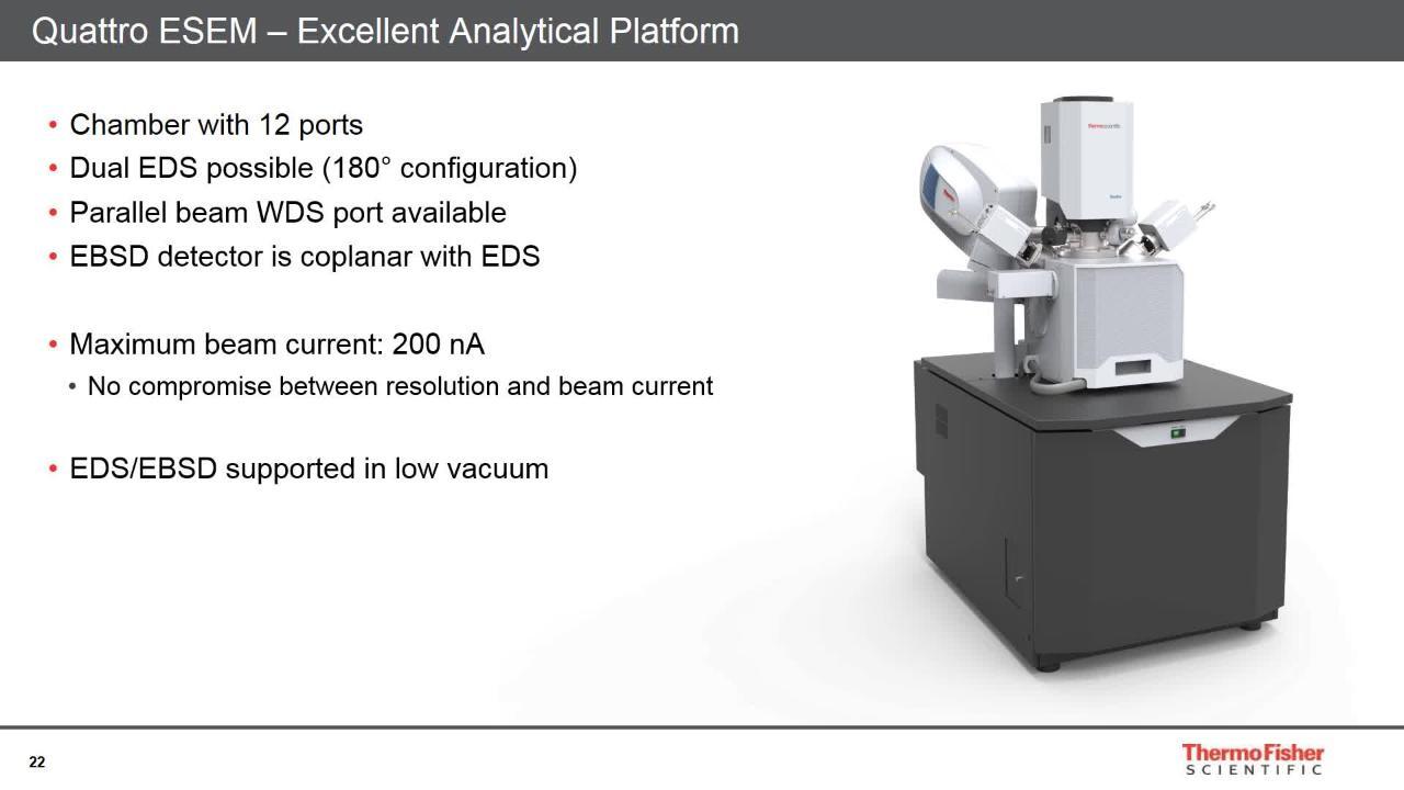
Thanks to the Quattro ESEM chamber size, it accommodates a wide range of accessories. Analytical capabilities include energy dispersive X-ray spectroscopy (EDS) with ports for 180-degree dual EDS attachment, electron backscatter diffraction (EBSD) coplanar with EDS, and wavelength dispersive X-ray spectroscopy (WDS). Furthermore, the Quattro ESEM comes with the new Thermo Scientific ChemiSEM Technology, a unique live elemental imaging capability that is fully integrated into the SEM user interface, providing always-available compositional information through the most intuitive interface.
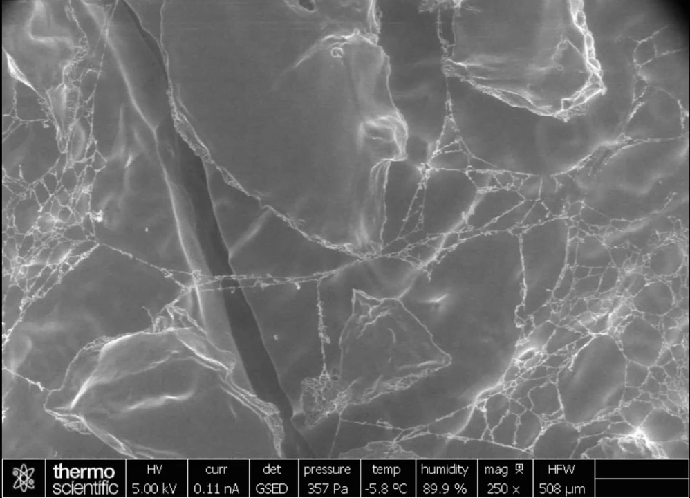
Diverse material application observed with the Quattro ESEM
Click image to enlarge
Metals and alloys (metallic filter).
Click image to enlarge
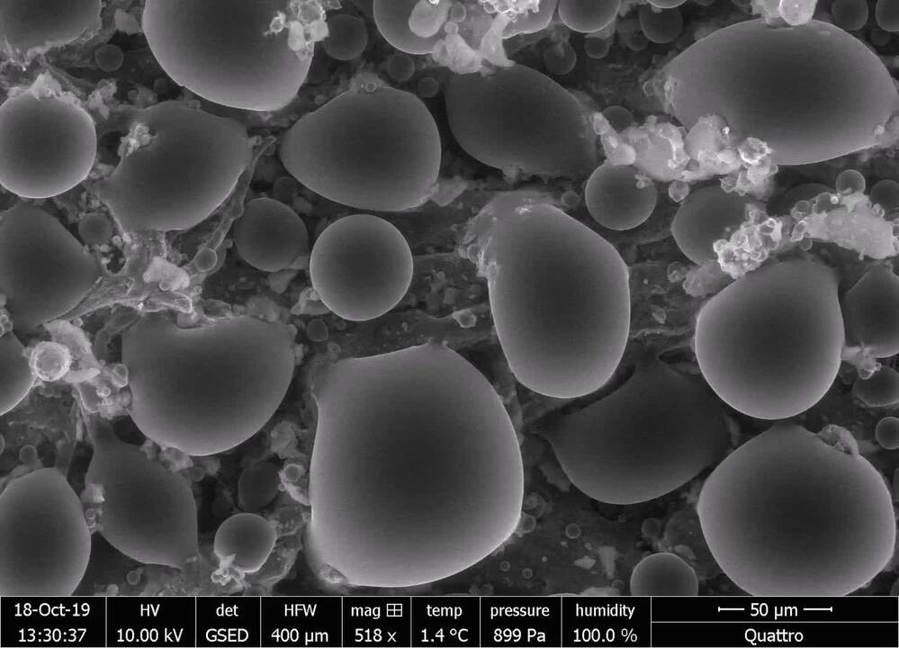
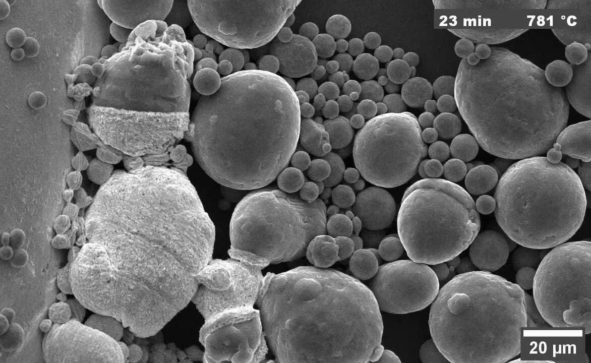
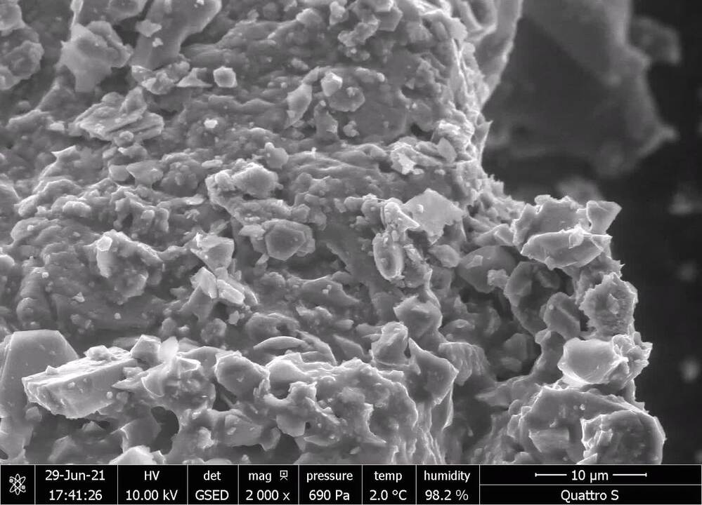
Additive manufacturing powder.
Click image to enlarge
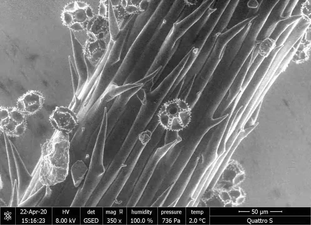
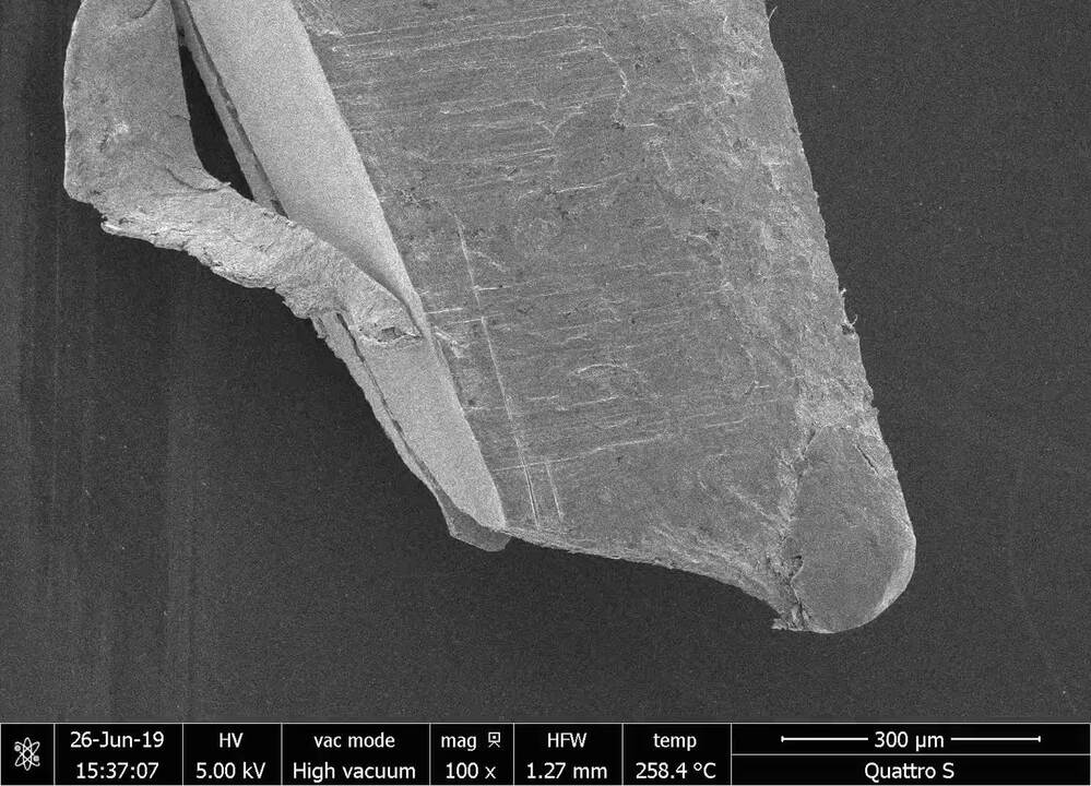
Soft materials (polymeric fibers).
Click image to enlarge
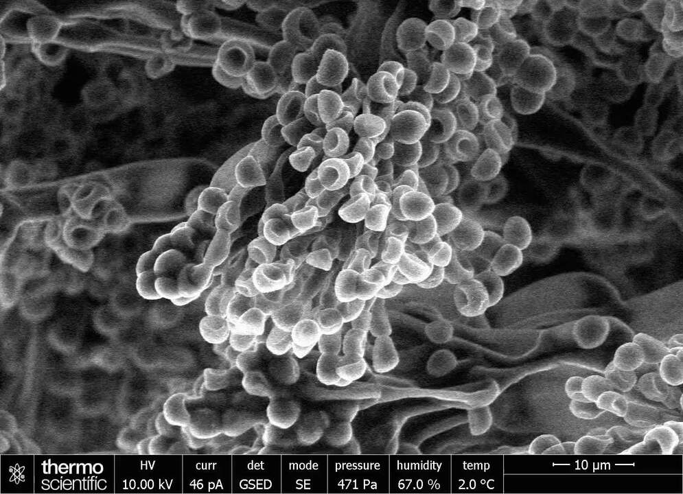
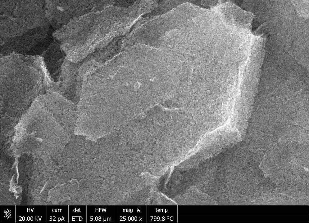
Biological samples (mold spores).
Click image to enlarge
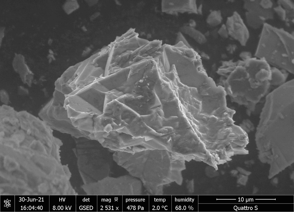
Geological materials (diamonds).
Click image to enlarge
Minerals (shell’s surface).
Click image to enlarge
Ceramics (zirconia in a thermal barrier coating).
Click image to enlarge
Soft materials (cross section of a tire).
Click image to enlarge
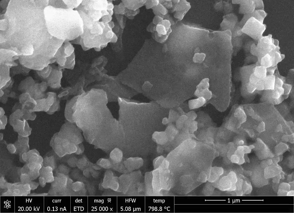
Chemicals (zinc oxide nanoparticles assembled into platelets).
Quattro Environmental Scanning Electron Microscope features
Dynamic in situ experiments
In situ study of materials in their natural state: unique high-resolution FEG-SEM with environmental mode (ESEM). In situ analysis at temperatures ranging from -165°C to 1,400°C with a range of cryo, Peltier, and heating stages.
Video Player is loading.
Current Time 0:00
/
Duration 0:17
Loaded: 55.66%
0:00
Stream Type LIVE
Remaining Time -0:17
1x
Chapters
descriptions off, selected
captions settings, opens captions settings dialog
captions off, selected
default, selected
This is a modal window.
Beginning of dialog window. Escape will cancel and close the window.
End of dialog window.
This is a modal window. This modal can be closed by pressing the Escape key or activating the close button.
This is a modal window. This modal can be closed by pressing the Escape key or activating the close button.
Peltier cooling experiment of sodium sulfate crystals in sandstone.
In situ cooling experiment conducted on a sandstone sample with crystals of sodium sulfate. The target of the experiment was to study the sodium sulfate behavior when completely hydrated. The pressure was varied up to 700 Pa, maintaining the temperature at 2˚C. Sample courtesy of Institute of Theoretical and Applied Mechanics of the Academy of Science, Czech Republic.
Video Player is loading.
Current Time 0:00
/
Duration 0:33
Loaded: 29.61%
0:00
Stream Type LIVE
Remaining Time -0:33
1x
Chapters
descriptions off, selected
captions settings, opens captions settings dialog
captions off, selected
default, selected
This is a modal window.
Beginning of dialog window. Escape will cancel and close the window.
End of dialog window.
This is a modal window. This modal can be closed by pressing the Escape key or activating the close button.
This is a modal window. This modal can be closed by pressing the Escape key or activating the close button.
μHeater Holder heating experiment of magnetite and hematite.
Heating experiment run with the μHeater Holder. The video shows the heating of a mix of magnetite and hematite from 40˚C up to 1,000˚C.
Wide range of information
Observe all information from all samples with simultaneous SE and BSE imaging in every mode of operation. Several additional detectors, such as the STEM3+ detector and the retractable RGB cathodoluminescence (CL) detector are available to accommodate every user’s need and provide a complete set of information from a wide range of materials.
Click image to enlarge
Material contrast from a mix of different metals.
Click image to enlarge
Bright field (BF) STEM3+ image of a biological section of a planarian.
Click image to enlarge
CL image of diamonds.
Excellent analytical capabilities
Excellent analytical capabilities with a flexible chamber that allows multiple EDS, EBSD, or WDS detectors. Elemental information at your fingertips with ChemiSEM Technology, which provides live, quantitative, elemental mapping for unprecedented time to result and ease of use. Excellent analysis of non-conductive samples: accurate EDS and EBSD are enabled in low vacuum with the Quattro ESEM's through-the-lens pumping.
Click image to enlarge
ChemiSEM image of dental filling material, showing a mixture of Hg, Cu, Sn and Ag.
Click image to enlarge
EBSD on ceramic materials in low vacuum.
Minimize sample preparation time with the unique combination of high-vacuum, low-vacuum, and environmental modes
Click image to enlarge
Polymeric fibers (low vacuum imaging, 80 Pa).
Click image to enlarge
Hydrated mold spores (ESEM imaging, 800 Pa).
Click image to enlarge
Salts on bacon (ESEM imaging, 700 Pa).
Ease of use with innovative options and advanced automation
Easy to use, intuitive software with User Guidance. The unique Undo function permits efficient exploration of imaging conditions and allows you to work faster with fewer mouse clicks.
Video Player is loading.
Current Time 0:00
/
Duration 0:30
Loaded: 32.52%
0:00
Stream Type LIVE
Remaining Time -0:30
1x
Chapters
descriptions off, selected
captions settings, opens captions settings dialog
captions off, selected
en (Main), selected
This is a modal window.
Beginning of dialog window. Escape will cancel and close the window.
End of dialog window.
This is a modal window. This modal can be closed by pressing the Escape key or activating the close button.
This is a modal window. This modal can be closed by pressing the Escape key or activating the close button.
Optimize your work and feel free to explore the imaging condition with the unique Undo function.
Advanced automation
Advanced automation is provided in different ways, with either Thermo Scientific AutoScript 4 Software or Thermo Scientific Maps Software, depending on the specific application needs.
AutoScript 4 Software offers control of the Quattro ESEM. Take advantage of a Python-based application programming interface (API) to optimize your in situ cooling or heating experiments or record and monitor dynamic parameters such as temperature, stage position, or pressure.
Maps Software automates large-area acquisition on multiple samples with up to four different simultaneous signals. In addition to a clear increase in the system productivity, it offers a multi-scale, multi-layered visualization environment in which 2D and 3D data and imagery can be imported from any source to correlate different modalities.
Video Player is loading.
Current Time 0:00
/
Duration 0:08
Loaded: 100.00%
0:00
Stream Type LIVE
Remaining Time -0:08
1x
Chapters
descriptions off, selected
captions settings, opens captions settings dialog
captions off, selected
default, selected
This is a modal window.
Beginning of dialog window. Escape will cancel and close the window.
End of dialog window.
This is a modal window. This modal can be closed by pressing the Escape key or activating the close button.
This is a modal window. This modal can be closed by pressing the Escape key or activating the close button.
Live drift compensation with AutoScript 4 Software on an ESEM heating stage experiment.
ESEM heating stage experiment on a silver wire, heated from 300˚C to 530˚C. AutoScript 4 Software was used to obtain a live drift compensation through a combination of beam shift and stage moves. The result was a stable image of the silver wire for the whole duration of the movie, even when the wire changed location over the substrate.
Specifications
Style Sheet for Products Table Specifications
Resolution
High-vacuum imaging
0.8 nm at 30 kV (STEM)
1.0 nm at 30 kV (SE) in high vacuum
1.3 nm at 30 kV (SE) in low vacuum and ESEM mode
3.0 nm at 1 kV (SE)
Standard detectors
ETD, low-vacuum SED (LVD), gaseous SED for ESEM mode (GSED), IR camera
Live quantitative SEM image coloring is available based on energy-dispersive X-ray spectroscopy (EDS). Point & ID, linescan, region, element maps and accurate Noran quantification are included.
Stage bias (beam deceleration, optional)
-4000 V to +50 V
Low vacuum mode
Up to 2600 Pa (H2O) or 4000 Pa (N2)
Stage
5-axis motorized eucentric stage, 110 x 110 mm2 with a 105° tilt range. Maximum sample weight: 5 kg in un-tilted position.
Standard sample holder
Standard multi-sample SEM holder uniquely mounts directly onto the stage, hosts up to 18 standard stubs (⌀ 12 mm), does not require tools to mount a sample
Chamber
340 mm inside width, 12 ports, three simultaneous EDS detectors possible, two at 180°, coplanar EDS/EBSD orthogonal to the tilt axis of the stage
In situ accessories (optional)
Software controlled -20° C to +60° C Peltier cold stage
Software controlled 1000° C low vacuum/ESEM heating stage
Software controlled 1100° C high vacuum heating stage
Software controlled 1400° C low vacuum/ESEM heating stage
Integrated gas injection: up to 2 units (other accessories may limit number of GIS available) for beam-induced deposition of the following materials:
Platinum
Tungsten
Carbon
Manipulators
Cryo-stage
Electrical probing / multi-probing stations
Software options
Maps Software for automatic large area acquisition using tiling and stitching; correlative work
CaCu3Ti4O12 (CCTO) is a compound with an extraordinarily high dielectric constant. Sample courtesy of Mr Sylvain Marinel, CRISMAT laboratory, France.
Click image to enlarge
The Quattro ESEM allows the study of samples at 100% humidity, such as these pollen grains, in ESEM mode.
Click image to enlarge
Salt crystals dissolving and recrystallizing as water is either condensing or evaporating in ESEM mode.
Click image to enlarge
Bright field (BF) STEM3+ image of a biological section of a planarian.
Click image to enlarge
Ceramic in glass.
Click image to enlarge
Ceramics and metals from a thermal barrier coating.
Click image to enlarge
ChemiSEM image of dental filling material.
Click image to enlarge
Coated microdrill.
Click image to enlarge
Crystals and surface details on rocks.
Click image to enlarge
Crystals and surface details on rocks.
Click image to enlarge
Crystals and surface details on rocks.
Click image to enlarge
Details of a butterfly wing.
Click image to enlarge
Details of a spider.
Click image to enlarge
CL image of diamonds.
Click image to enlarge
Diatom.
Click image to enlarge
Glass fibers.
Click image to enlarge
Iron oxide.
Click image to enlarge
Metallic filter.
Click image to enlarge
Mix of different metals.
Click image to enlarge
Material contrast from a mix of different metals.
Click image to enlarge
Biological samples (mold spores).
Click image to enlarge
Polymeric fibers.
Click image to enlarge
Red, green, blue emitting particles from a cathode tube.
Click image to enlarge
Tin dendrites.
Tin dendrites.
Click image to enlarge
Worn textile fabric.
Click image to enlarge
Zircons.
Video Player is loading.
Current Time 0:00
/
Duration 32:12
Loaded: 0.51%
00:00
Stream Type LIVE
Remaining Time -32:12
1x
Chapters
descriptions off, selected
captions settings, opens captions settings dialog
captions off, selected
en (Main), selected
This is a modal window.
Beginning of dialog window. Escape will cancel and close the window.
End of dialog window.
This is a modal window. This modal can be closed by pressing the Escape key or activating the close button.
This is a modal window. This modal can be closed by pressing the Escape key or activating the close button.
Video Player is loading.
Current Time 0:00
/
Duration 0:08
Loaded: 100.00%
0:00
Stream Type LIVE
Remaining Time -0:08
1x
Chapters
descriptions off, selected
captions settings, opens captions settings dialog
captions off, selected
default, selected
This is a modal window.
Beginning of dialog window. Escape will cancel and close the window.
End of dialog window.
This is a modal window. This modal can be closed by pressing the Escape key or activating the close button.
This is a modal window. This modal can be closed by pressing the Escape key or activating the close button.
ESEM cooling experiment on coated filter paper, showing the paper’s behavior when fully hydrated.
Video Player is loading.
Current Time 0:00
/
Duration 0:37
Loaded: 26.76%
0:00
Stream Type LIVE
Remaining Time -0:37
1x
Chapters
descriptions off, selected
captions settings, opens captions settings dialog
captions off, selected
default, selected
This is a modal window.
Beginning of dialog window. Escape will cancel and close the window.
End of dialog window.
This is a modal window. This modal can be closed by pressing the Escape key or activating the close button.
This is a modal window. This modal can be closed by pressing the Escape key or activating the close button.
WetSTEM cooling experiment on a flower and pollen sample. Thanks to the flexible design of this stage, it allows the characterization both in transmission mode and in top-down mode.
Video Player is loading.
Current Time 0:00
/
Duration 0:58
Loaded: 17.04%
0:00
Stream Type LIVE
Remaining Time -0:58
1x
Chapters
descriptions off, selected
captions settings, opens captions settings dialog
captions off, selected
default, selected
This is a modal window.
Beginning of dialog window. Escape will cancel and close the window.
End of dialog window.
This is a modal window. This modal can be closed by pressing the Escape key or activating the close button.
This is a modal window. This modal can be closed by pressing the Escape key or activating the close button.
Peltier cooling experiment on food gelatin. The video shows the microstructural changes of the gelatine when frozen down to -10˚C.
Video Player is loading.
Current Time 0:00
/
Duration 0:33
Loaded: 29.61%
0:00
Stream Type LIVE
Remaining Time -0:33
1x
Chapters
descriptions off, selected
captions settings, opens captions settings dialog
captions off, selected
default, selected
This is a modal window.
Beginning of dialog window. Escape will cancel and close the window.
End of dialog window.
This is a modal window. This modal can be closed by pressing the Escape key or activating the close button.
This is a modal window. This modal can be closed by pressing the Escape key or activating the close button.
Heating experiment ran with the μHeater system. The video shows the heating of a mix of magnetite and hematite from 40˚C up to 1000˚C.
Video Player is loading.
Current Time 0:00
/
Duration 1:13
Loaded: 13.60%
0:00
Stream Type LIVE
Remaining Time -1:13
1x
Chapters
descriptions off, selected
captions settings, opens captions settings dialog
captions off, selected
default, selected
This is a modal window.
Beginning of dialog window. Escape will cancel and close the window.
End of dialog window.
This is a modal window. This modal can be closed by pressing the Escape key or activating the close button.
This is a modal window. This modal can be closed by pressing the Escape key or activating the close button.
Metals heating with the high vacuum heating stage. the video shows how the melted gold moves over the other materials while the heating progresses.
Video Player is loading.
Current Time 0:00
/
Duration 0:26
Loaded: 37.08%
0:00
Stream Type LIVE
Remaining Time -0:26
1x
Chapters
descriptions off, selected
captions settings, opens captions settings dialog
captions off, selected
default, selected
This is a modal window.
Beginning of dialog window. Escape will cancel and close the window.
End of dialog window.
This is a modal window. This modal can be closed by pressing the Escape key or activating the close button.
This is a modal window. This modal can be closed by pressing the Escape key or activating the close button.
Cooling experiment conducted with the Peltier cooling stage. It shows the hydration of mold spores from a cheese sample.
Video Player is loading.
Current Time 0:00
/
Duration 0:17
Loaded: 55.66%
0:00
Stream Type LIVE
Remaining Time -0:17
1x
Chapters
descriptions off, selected
captions settings, opens captions settings dialog
captions off, selected
default, selected
This is a modal window.
Beginning of dialog window. Escape will cancel and close the window.
End of dialog window.
This is a modal window. This modal can be closed by pressing the Escape key or activating the close button.
This is a modal window. This modal can be closed by pressing the Escape key or activating the close button.
In-situ cooling experiment conducted on a sandstone sample with crystals of sodium sulphate, to study its behavior when completely hydrated. Sample courtesy of Institute of Theoretical and Applied Mechanics of the Academy of Science, Czech Republic.
Video Player is loading.
Current Time 0:00
/
Duration 0:25
Loaded: 38.96%
0:00
Stream Type LIVE
Remaining Time -0:25
1x
Chapters
descriptions off, selected
captions settings, opens captions settings dialog
captions off, selected
default, selected
This is a modal window.
Beginning of dialog window. Escape will cancel and close the window.
End of dialog window.
This is a modal window. This modal can be closed by pressing the Escape key or activating the close button.
This is a modal window. This modal can be closed by pressing the Escape key or activating the close button.
In-situ cooling experiment conducted on a sandstone sample with crystals of sodium sulphate, to study its behavior when completely hydrated. Sample courtesy of Institute of Theoretical and Applied Mechanics of the Academy of Science, Czech Republic.
Video Player is loading.
Current Time 0:00
/
Duration 0:08
Loaded: 100.00%
0:00
Stream Type LIVE
Remaining Time -0:08
1x
Chapters
descriptions off, selected
captions settings, opens captions settings dialog
captions off, selected
default, selected
This is a modal window.
Beginning of dialog window. Escape will cancel and close the window.
End of dialog window.
This is a modal window. This modal can be closed by pressing the Escape key or activating the close button.
This is a modal window. This modal can be closed by pressing the Escape key or activating the close button.
ESEM heating stage experiment on a silver wire, heated from 300˚C to 530˚C. Autoscript 4 was used to obtain a live drift compensation through a combination of beam shift and stage moves.
Video Player is loading.
Current Time 0:00
/
Duration 0:14
Loaded: 67.04%
0:00
Stream Type LIVE
Remaining Time -0:14
1x
Chapters
descriptions off, selected
captions settings, opens captions settings dialog
captions off, selected
default, selected
This is a modal window.
Beginning of dialog window. Escape will cancel and close the window.
End of dialog window.
This is a modal window. This modal can be closed by pressing the Escape key or activating the close button.
This is a modal window. This modal can be closed by pressing the Escape key or activating the close button.
Heating of a solder wire from 200˚C to 350˚C ran with the high vacuum heating stage.
Video Player is loading.
Current Time 0:00
/
Duration 0:19
Loaded: 52.08%
0:00
Stream Type LIVE
Remaining Time -0:19
1x
Chapters
descriptions off, selected
captions settings, opens captions settings dialog
captions off, selected
default, selected
This is a modal window.
Beginning of dialog window. Escape will cancel and close the window.
End of dialog window.
This is a modal window. This modal can be closed by pressing the Escape key or activating the close button.
This is a modal window. This modal can be closed by pressing the Escape key or activating the close button.
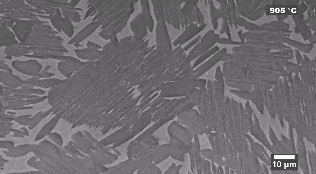
TiAl TNM-B1 alloy heating with the high vacuum heating stage. The alloy was heated from room temperature up to 1100˚C showing oxidation and phase changes on the surface.
Video Player is loading.
Current Time 0:00
/
Duration 0:35
Loaded: 27.78%
0:00
Stream Type LIVE
Remaining Time -0:35
1x
Chapters
descriptions off, selected
captions settings, opens captions settings dialog
captions off, selected
default, selected
This is a modal window.
Beginning of dialog window. Escape will cancel and close the window.
End of dialog window.
This is a modal window. This modal can be closed by pressing the Escape key or activating the close button.
This is a modal window. This modal can be closed by pressing the Escape key or activating the close button.
μHeater heating experiment on zinc oxide platelets. The progressive heating up to 1000˚C shows the textural changes of the platelet.
Webinar: Scanning electron microscopy: selecting the right technology for your needs
This on-demand webinar has been designed to help you decide which SEM best meets your unique needs. We present an overview of Thermo Fisher Scientific SEM technology for multi-user research labs and focus on how these wide-ranging solutions deliver performance, versatility, in situ dynamics and faster time to results. Watch this webinar if you are interested in:
How the needs for different microanalysis modalities are met (EDX, EBSD, WDS, CL, etc.).
How samples are characterized in their natural state without the need for sample preparation.
How new advanced automation allows researchers to save time and increase productivity.
Beginning of dialog window. Escape will cancel and close the window.
End of dialog window.
This is a modal window. This modal can be closed by pressing the Escape key or activating the close button.
This is a modal window. This modal can be closed by pressing the Escape key or activating the close button.
Video Player is loading.
Current Time 0:00
/
Duration 0:08
Loaded: 100.00%
0:00
Stream Type LIVE
Remaining Time -0:08
1x
Chapters
descriptions off, selected
captions settings, opens captions settings dialog
captions off, selected
default, selected
This is a modal window.
Beginning of dialog window. Escape will cancel and close the window.
End of dialog window.
This is a modal window. This modal can be closed by pressing the Escape key or activating the close button.
This is a modal window. This modal can be closed by pressing the Escape key or activating the close button.
ESEM cooling experiment on coated filter paper, showing the paper’s behavior when fully hydrated.
Video Player is loading.
Current Time 0:00
/
Duration 0:37
Loaded: 26.76%
0:00
Stream Type LIVE
Remaining Time -0:37
1x
Chapters
descriptions off, selected
captions settings, opens captions settings dialog
captions off, selected
default, selected
This is a modal window.
Beginning of dialog window. Escape will cancel and close the window.
End of dialog window.
This is a modal window. This modal can be closed by pressing the Escape key or activating the close button.
This is a modal window. This modal can be closed by pressing the Escape key or activating the close button.
WetSTEM cooling experiment on a flower and pollen sample. Thanks to the flexible design of this stage, it allows the characterization both in transmission mode and in top-down mode.
Video Player is loading.
Current Time 0:00
/
Duration 0:58
Loaded: 17.04%
0:00
Stream Type LIVE
Remaining Time -0:58
1x
Chapters
descriptions off, selected
captions settings, opens captions settings dialog
captions off, selected
default, selected
This is a modal window.
Beginning of dialog window. Escape will cancel and close the window.
End of dialog window.
This is a modal window. This modal can be closed by pressing the Escape key or activating the close button.
This is a modal window. This modal can be closed by pressing the Escape key or activating the close button.
Peltier cooling experiment on food gelatin. The video shows the microstructural changes of the gelatine when frozen down to -10˚C.
Video Player is loading.
Current Time 0:00
/
Duration 0:33
Loaded: 29.61%
0:00
Stream Type LIVE
Remaining Time -0:33
1x
Chapters
descriptions off, selected
captions settings, opens captions settings dialog
captions off, selected
default, selected
This is a modal window.
Beginning of dialog window. Escape will cancel and close the window.
End of dialog window.
This is a modal window. This modal can be closed by pressing the Escape key or activating the close button.
This is a modal window. This modal can be closed by pressing the Escape key or activating the close button.
Heating experiment ran with the μHeater system. The video shows the heating of a mix of magnetite and hematite from 40˚C up to 1000˚C.
Video Player is loading.
Current Time 0:00
/
Duration 1:13
Loaded: 13.60%
0:00
Stream Type LIVE
Remaining Time -1:13
1x
Chapters
descriptions off, selected
captions settings, opens captions settings dialog
captions off, selected
default, selected
This is a modal window.
Beginning of dialog window. Escape will cancel and close the window.
End of dialog window.
This is a modal window. This modal can be closed by pressing the Escape key or activating the close button.
This is a modal window. This modal can be closed by pressing the Escape key or activating the close button.
Metals heating with the high vacuum heating stage. the video shows how the melted gold moves over the other materials while the heating progresses.
Video Player is loading.
Current Time 0:00
/
Duration 0:26
Loaded: 37.08%
0:00
Stream Type LIVE
Remaining Time -0:26
1x
Chapters
descriptions off, selected
captions settings, opens captions settings dialog
captions off, selected
default, selected
This is a modal window.
Beginning of dialog window. Escape will cancel and close the window.
End of dialog window.
This is a modal window. This modal can be closed by pressing the Escape key or activating the close button.
This is a modal window. This modal can be closed by pressing the Escape key or activating the close button.
Cooling experiment conducted with the Peltier cooling stage. It shows the hydration of mold spores from a cheese sample.
Video Player is loading.
Current Time 0:00
/
Duration 0:17
Loaded: 55.66%
0:00
Stream Type LIVE
Remaining Time -0:17
1x
Chapters
descriptions off, selected
captions settings, opens captions settings dialog
captions off, selected
default, selected
This is a modal window.
Beginning of dialog window. Escape will cancel and close the window.
End of dialog window.
This is a modal window. This modal can be closed by pressing the Escape key or activating the close button.
This is a modal window. This modal can be closed by pressing the Escape key or activating the close button.
In-situ cooling experiment conducted on a sandstone sample with crystals of sodium sulphate, to study its behavior when completely hydrated. Sample courtesy of Institute of Theoretical and Applied Mechanics of the Academy of Science, Czech Republic.
Video Player is loading.
Current Time 0:00
/
Duration 0:25
Loaded: 38.96%
0:00
Stream Type LIVE
Remaining Time -0:25
1x
Chapters
descriptions off, selected
captions settings, opens captions settings dialog
captions off, selected
default, selected
This is a modal window.
Beginning of dialog window. Escape will cancel and close the window.
End of dialog window.
This is a modal window. This modal can be closed by pressing the Escape key or activating the close button.
This is a modal window. This modal can be closed by pressing the Escape key or activating the close button.
In-situ cooling experiment conducted on a sandstone sample with crystals of sodium sulphate, to study its behavior when completely hydrated. Sample courtesy of Institute of Theoretical and Applied Mechanics of the Academy of Science, Czech Republic.
Video Player is loading.
Current Time 0:00
/
Duration 0:08
Loaded: 62.50%
0:00
Stream Type LIVE
Remaining Time -0:08
1x
Chapters
descriptions off, selected
captions settings, opens captions settings dialog
captions off, selected
default, selected
This is a modal window.
Beginning of dialog window. Escape will cancel and close the window.
End of dialog window.
This is a modal window. This modal can be closed by pressing the Escape key or activating the close button.
This is a modal window. This modal can be closed by pressing the Escape key or activating the close button.
ESEM heating stage experiment on a silver wire, heated from 300˚C to 530˚C. Autoscript 4 was used to obtain a live drift compensation through a combination of beam shift and stage moves.
Video Player is loading.
Current Time 0:00
/
Duration 0:14
Loaded: 53.63%
0:00
Stream Type LIVE
Remaining Time -0:14
1x
Chapters
descriptions off, selected
captions settings, opens captions settings dialog
captions off, selected
default, selected
This is a modal window.
Beginning of dialog window. Escape will cancel and close the window.
End of dialog window.
This is a modal window. This modal can be closed by pressing the Escape key or activating the close button.
This is a modal window. This modal can be closed by pressing the Escape key or activating the close button.
Heating of a solder wire from 200˚C to 350˚C ran with the high vacuum heating stage.
Video Player is loading.
Current Time 0:00
/
Duration 0:19
Loaded: 0.00%
0:00
Stream Type LIVE
Remaining Time -0:19
1x
Chapters
descriptions off, selected
captions settings, opens captions settings dialog
captions off, selected
default, selected
This is a modal window.
Beginning of dialog window. Escape will cancel and close the window.
End of dialog window.
This is a modal window. This modal can be closed by pressing the Escape key or activating the close button.
This is a modal window. This modal can be closed by pressing the Escape key or activating the close button.
TiAl TNM-B1 alloy heating with the high vacuum heating stage. The alloy was heated from room temperature up to 1100˚C showing oxidation and phase changes on the surface.
Video Player is loading.
Current Time 0:00
/
Duration 0:35
Loaded: 0.00%
0:00
Stream Type LIVE
Remaining Time -0:35
1x
Chapters
descriptions off, selected
captions settings, opens captions settings dialog
captions off, selected
default, selected
This is a modal window.
Beginning of dialog window. Escape will cancel and close the window.
End of dialog window.
This is a modal window. This modal can be closed by pressing the Escape key or activating the close button.
This is a modal window. This modal can be closed by pressing the Escape key or activating the close button.
μHeater heating experiment on zinc oxide platelets. The progressive heating up to 1000˚C shows the textural changes of the platelet.
Webinar: Scanning electron microscopy: selecting the right technology for your needs
This on-demand webinar has been designed to help you decide which SEM best meets your unique needs. We present an overview of Thermo Fisher Scientific SEM technology for multi-user research labs and focus on how these wide-ranging solutions deliver performance, versatility, in situ dynamics and faster time to results. Watch this webinar if you are interested in:
How the needs for different microanalysis modalities are met (EDX, EBSD, WDS, CL, etc.).
How samples are characterized in their natural state without the need for sample preparation.
How new advanced automation allows researchers to save time and increase productivity.
Modern industry demands high throughput with superior quality, a balance that is maintained through robust process control. SEM and TEM tools with dedicated automation software provide rapid, multi-scale information for process monitoring and improvement.
Quality control and assurance are essential in modern industry. We offer a range of EM and spectroscopy tools for multi-scale and multi-modal analysis of defects, allowing you to make reliable and informed decisions for process control and improvement.
Novel materials are investigated at increasingly smaller scales for maximum control of their physical and chemical properties. Electron microscopy provides researchers with key insight into a wide variety of material characteristics at the micro- to nano-scale.
Innovation starts with research and development. Learn more about solutions to help you understand innovative structures and materials at the atomic level.
Manufacturing today’s complex semiconductors requires exact process controls. Learn more about advanced metrology and analysis solutions to accelerate yield learnings.
Complex semiconductor device structures result in more places for defects to hide. Learn more about failure analysis solutions to isolate, analyze, and repair defects.
Energy dispersive spectroscopy (EDS) collects detailed elemental information along with electron microscopy images, providing critical compositional context for EM observations. With EDS, chemical composition can be determined from quick, holistic surface scans down to individual atoms.
Studying materials in real-world conditions often involves working at high temperatures. The behavior of materials as they recrystallize, melt, deform, or react in the presence of heat can be studied in situ with scanning electron microscopy or DualBeam tools.
Environmental SEM allows materials to be imaged in their native state. This is ideally suited for academic and industrial researchers who need to test and analyze samples that are wet, dirty, reactive, outgassing or otherwise not vacuum compatible.
Direct, real-time observation of microstructural changes with electron microscopy is necessary to understand the underlying principles of dynamic processes such as recrystallization, grain growth, and phase transformation during heating, cooling, and wetting.
Particle analysis plays a vital role in nanomaterials research and quality control. The nanometer-scale resolution and superior imaging of electron microscopy can be combined with specialized software for rapid characterization of powders and particles.
Cathodoluminescence (CL) describes the emission of light from a material when it is excited by an electron beam. This signal, captured by a specialized CL detector, carries information on the sample’s composition, crystal defects, or photonic properties.
Thermo Fisher Scientific offers scanning electron microscopes for every function of a semiconductor lab, from general imaging tasks to advanced failure analysis techniques requiring precise voltage-contrast measurements.
Style Sheet to change H2 style to p with em-h2-header class
Contact us
Electron microscopy services
To ensure optimal system performance, we provide you access to a world-class network of field service experts, technical support, and certified spare parts.

")


 is a compound with an extraordinarily high dielectric constant. Sample courtesy of Mr Sylvain Marinel, CRISMAT laboratory, France.")


.")


_Technique_800x375_144DPI.jpg)


