Mechanism of Ebola cell entry is critical for the development of effective therapeutics
The Ebola virus is a notorious illness for both its high contagiousness and lethality, marked by dramatic external bleeding in its later stages. In recent years, there has been great progress in the development of preventative treatments against Ebola and an FDA approved vaccine has been available since 2019. However, the finer mechanistic behavior of Ebola and the Ebola virus structure remains relatively unknown, which hampers the development of treatments and therapeutics. In particular, capturing the mechanism of Ebola cell entry has been difficult, as it is a highly dynamic process which ends with the delivering of the Ebola virus RNA into the host cell. Researchers from the University Hospital Heidelberg (Germany) have now visualized Ebola virus membrane fusion and cell entry through a combination of cryo-electron tomography (cryo-ET) imaging, digital 3D reconstruction, molecular analysis and membrane modeling.
Determining Ebola virus structure and cell entry
Ebola viruses (EBOVs) form filamentous and long virions which enter into the cytoplasm of the host cell through the fusion with endosomal membranes. A matrix layer consisting of the matrix viral protein 40 (VP40) is fundamental to determine the shape and the stability of the EBOVs and forms a scaffold under the viral envelope which is important to protect the genome during the transmission. However, it remains unknown how virions go through the uncoating process, including virion disassembly and release of the nucleocapsid, (which contains the viral genome) into the cytosol. Using cryo-ET, researchers first produce a thin slice of a flash-frozen biological sample with a dedicated cryo-focused ion beam (cryo-FIB), which can mill a specimen down to nanometer thickness. Then, the sample is imaged with a cryo-ET microscope (in 2D) at a series of angles, which are digitally reconstructed into a 3D representation of the sample.
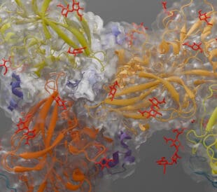
In this study, tomograms acquired with Thermo Fisher Scientific Titan Krios showed impressive details of the EBOV virions localized in the endosomes of infected cells (Figure 1). The 3D segmentation and reconstruction of tomograms carried out with Thermo Scientific Amira Software provided incredible details of viral and cellular structures. Researchers visualized membrane fragments and vesicles, which were products of a degradation process that occurred in the host cell. The VP40 matrix appeared disassembled and not attached to the viral envelope as well. Interestingly Amira Software allowed to distinguish key components of the Ebola virus structure such as the envelope, VP40 and the nucleocapsid.
By analyzing multiple endosomes and combining their observations with molecular analysis and membrane modeling, they were able to determine that the disassembly of the VP40 matrix occurs at low pH conditions prior to the membrane fusion. The researchers suggest that the EBOV uncoating starts with the disassembly of the VP40 matrix followed by the membrane fusion and release of the nucleocapsid into the cytoplasm.

Cryo-ET process along with resulting 2D images and 3D reconstructions. Segmentation performed with Amira Software clearly distinguishes key components of the Ebola virion. Figure reproduced in part from S.L. Winter, et al. under CC BY 4.0.
Producing targeted treatments by observing how viruses enter cells
Deconvoluting the way viruses enter our cells is a critical step towards targeted treatments. The behavior of the protein matrix revealed by the team at the University Hospital Heidelberg has provided a promising target for disrupting Ebola’s ability to infect cells. Their approach, which combines high-resolution cryo-EM with advanced segmentation and molecular modeling, serves as a novel template for the mechanistic investigation of any number of infectious diseases. We are excited to see how such direct observation of virus behavior will guide the development of effective treatments.
Learn more about Amira Software at thermofisher.com/amira >>
Rosa Pipitone PhD is a Marketing Content Specialist at Thermo Fisher Scientific. Written in collaboration with Alex Ilitchev PhD, Scientific Editor at Thermo Fisher Scientific.
Guiding the Development of an HIV Vaccine with Structure-Based Drug Design
Novel drug development in the fight against the HIV pandemic...
Read More
Virus microscopy unravels chikungunya virus structure
Global impacts of the chikungunya virus Chikungunya is a vir...
Read More
Finding new ways to treat pain with novel opioid receptor ligands
Analgesics, pain relief, and opioid addiction While pain man...
Read More
Glacios 2 Cryo-TEM and the Evolution of 200 kV Cryo-EM
Glacios 2 Cryo-TEM and the Evolution of 200 kV Cryo-EM Cryo-...
Read More
Yes