Ion channel structure
Ion channels are a broad category of protein complexes that serve as openings in a cell membrane, enabling and regulating the passage of various ionic species between the cell interior and the extracellular medium. These channels play an indispensable role in the maintenance and homeostasis of our cells. Dysfunction in ion channel behavior has been linked to an enormous variety of disorders (also called channelopathies) including cancers, dementia, diabetes, asthma and more. Despite this fact, ion channels prove to be a difficult target for drug design, primarily because selective inhibitor design is challenging and a lack of specificity can negatively impact other critical, structurally similar ion channels.

Cryo-EM map of the SARS-CoV-2 3a ion channel in a lipid nanodiscs at 2.9 Å nominal resolution, viewed from the membrane plane. Figure by David M. Kern et al. in Cryo-EM structure of the SARS-CoV-2 3a ion channel in lipid nanodiscs, available under CC BY 4.0.
Ion channel structure determination
As membrane proteins, ion channels pose several challenges to traditional structural analysis with techniques such as X-ray crystallography. For one, these complexes often rely (at least in part) on the membrane to maintain their structure and isolating them for crystallization can disrupt them from their native state. More critically, the different conformations of the ion channel are vital for therapeutic development, as they reveal the binding sites and transitional states of the ion channel. Any method that does not allow for the differentiation between these forms loses the necessary context for structure-based drug design.
Cryo-electron microscopy of ion channels
Cryo-electron microscopy (cryo-EM) describes a range of techniques including cryo-electron tomography, micro-electron diffraction, and single particle analysis. In the case of ion channels, cryo-EM has been used synonymously with single particle analysis of channel protein complexes embedded in nanodiscs. These lipid structures imitate the cell membrane and help to preserve the native state of the ion channel. Cryo-EM images thousands of individual nanodisc complexes from various angles and then combines these 2D images into a 3D reconstruction of the channel. Since the images can be categorized and grouped, the different conformations of the channel can be isolated and individually recreated from the same sample.
Cryo-EM in ion channel receptor analysis
The resolution and quality of cryo-EM is great enough to show the binding of agonist and antagonist molecules to the ion channel, revealing the precise location and conformation of potential therapeutic receptors. Below are just a few examples of how pharmaceutical companies have utilized the power of cryo-EM to study ion channels and advance structure-based drug design:
TRPC ion channel structure with cryo-EM
- Transient receptor potential-canonical (TRPC) proteins compose a family of non-specific cation channels associated with a variety of pathological conditions. Despite therapeutic potential, their structure and mechanism of action have remained largely unknown. Using cryo-EM, researchers at Amgen were not only able to obtain a high-resolution structure of TRPC6 but were also able to determine structures for the antagonist- and agonist-bound states. By identifying these recognition sites, Amgen is paving the way for future drug design against TRPC-related diseases.

Electron density map (A) and overlaid protein structure (B) of TRPC6 with bound agonist (magenta). Figure by Yonghong Bai, et al. in Structural basis for pharmacological modulation of the TRPC6 channel, available under CC BY 4.0.
Sodium ion channel structure with cryo-EM
- Voltage-gated sodium ion (Nav) channels are a critical area of pharmaceutical research as they are targets for toxins, therapeutic agents, and are susceptible to disease mutations. Notably, mutations to these channels have been associated with migraines, epilepsy, pain, as well as cardiac and muscle paralysis syndromes. The mechanistic behavior of these channels, however, continues to be poorly understood. Researchers at Genentech (Roche Group) utilized cryo-EM to study the association of Nav7 with Protoxin II (ProTx2), a channel antagonist isolated from the Peruvian green velvet tarantula. Researchers were able to determine the structure of both activated and inactivated Nav1.7, providing a crucial starting point for the development of specialized, therapeutic Nav antagonists.

Cryo-EM clearly shows the structural differences between the activated (blue/magenta) and deactivated (black) Nav1.7 ion channel bound to the ProTx2 antagonist. Image based on PDB entries 6N4R and 6N4Q, created with PyMOL.
PDB ion channel structure trends
The above results highlight the great potential of cryo-EM in resolving the structure and function of historically difficult membrane-bound proteins. The increasing number of ion channel structures determined with cryo-EM that appear in the Protein Data Bank clearly indicate the growing interest and utility of this technique. Pharmaceutical structure-based drug design is not just following this trend, but leading it, with novel results that are informing the next generation of therapeutics.
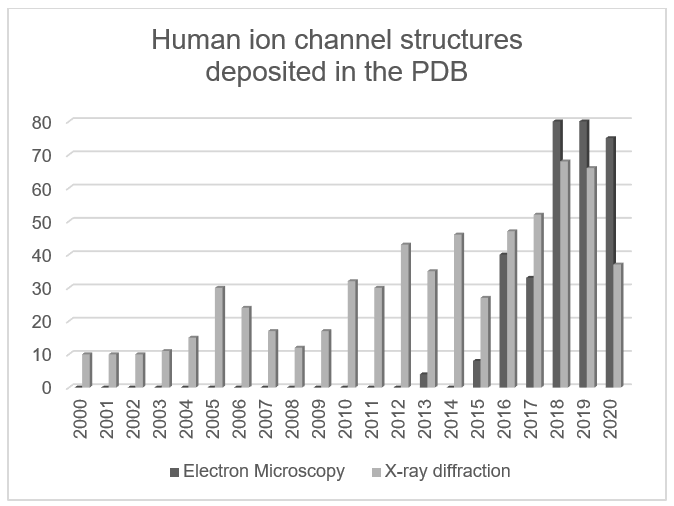
Source: Protein Data Bank (September 10, 2020)
Visit our Cryo-EM in the Pharmaceutical Industry webpage to learn more about how other pharmaceutical companies are using cryo-EM to accelerate their drug discovery.
To find out how you can incorporate cryo-EM into your pharmaceutical research, please see our Bio-pharmaceutical Research with Electron Microscopy page.
Hans Raaijmakers, PhD, is a Staff Scientist at Thermo Fisher Scientific.
///
Speak with an expert: https://www.thermofisher.com/blog/microscopy/speak-with-an-expert/
Subscribe now to receive Accelerating Microscopy updates straight to your inbox.

Decoding tauopathies: how cutting-edge cryo-EM is unraveling tau protein and Alzheimer’s disease
Tauopathy and neurodegeneration Neurodegenerative diseases a...
Read More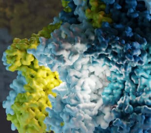
What is cryo-EM?
Cryo-EM: cryo electron microscopy Cryo-electron microscopy (...
Read More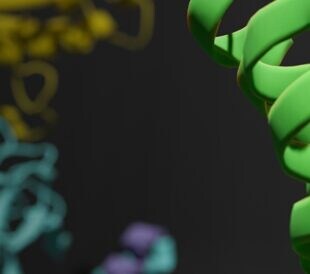
Finding new ways to treat pain with novel opioid receptor ligands
Analgesics, pain relief, and opioid addiction While pain man...
Read More
Glacios 2 Cryo-TEM and the Evolution of 200 kV Cryo-EM
Glacios 2 Cryo-TEM and the Evolution of 200 kV Cryo-EM Cryo-...
Read More
Leave a Reply