Throughout history, our world has been exposed to countless viruses and diseases. From the height of smallpox in the mid-1700s and polio in the 1950s to the present-day surge in Zika and Ebola outbreaks, vaccines and antiviral drugs have helped contain such ailments from spreading widely. However, there are still a number of viruses and diseases that still go untreated.
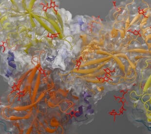
Cryo-electron microscopy (cryo-EM) is opening the door for researchers to determine how protein structures associated with more advanced viruses and diseases function and interact. In the last year alone, significant advances have been made:

Cryo-EM structures of tau filaments from Alzheimer’s disease via doi.org/10.1038/nature23002
Alzheimer’s
With the first electron microscope invented in 1931, not too long after the discovery of Alzheimer’s in 1906, leading scientists were soon able to study brain cells with even greater strength. While we still don’t know exactly what causes the neurodegenerative disease, scientists throughout history have leaned on progressive instruments like cryo-EM to supplement their research, leading to hundreds of new research and treatments. In fact, recent research conducted by scientists at the National Institute of Aging found new evidence on the viral species, noting the herpes viruses may play a role in Alzheimer’s disease biology.

Hydrophobic cleft at the growing end of α-Syn(1-121) fibrils via 10.7554/eLife.36402
Parkinson’s
As one of the most studied diseases in history, Parkinson’s – a disorder of the central nervous system that impacts movement and often results in tremors – has affected more than 10 million people worldwide to date. While no two people may have the same Parkinson’s experience, the disease is an extremely diverse disorder, including loss of dopaminergic neurons in the area of the brain known as the substantia nigra. Recent cryo-EM research from Vanderbilt University even suggests that deep brain stimulation administered to patients with early-stage Parkinson’s disease will slow the progression of rest tremor(s).

Structure of the HMPV N0-P complex via 10.7554/eLife.12627
Ebola
Our world recently watched the Ebola outbreak in West Africa – one of the most impactful since the discovery of the virus in 1976. As the virus known to cause haemorraghic fever and internal organ malfunction, Ebola has infected more than 10,000 individuals worldwide. Earlier this year, a group of researchers at major institutes such as Stanford, Washington University and Baylor used cryo-EM to image Ebola’s viral nucleocapsid-like assembly, which led to the proposal of how the virus may be targeted by antivirals.

The DENV2 and ZIKV E proteins share a highly similar fold and 54% sequence identity. Via 10.1073/pnas.1607931113
Zika
Aedes mosquito-borne disease, the Zika virus, was found in February of 2016 and, within a few months, the virus had infected more than one million people spread across more than 70 countries. In an effort to find treatments, research scientists recently began leveraging the latest electron microscopy techniques, leading to imaging of the highest-resolution image of the Zika virus to date. With the ability to now visualize the atomic details and stability of the virus’ structure in comparison to its flavivirus cousins, scientists at Purdue University have broken ground in designing vaccines and engineering anti-viral compounds that inhibit the virus.
In our upcoming series, we’ll be diving into the history and recent advancements of each disease to show how scientists are using cryo-EM to get one step closer to finding treatments and cures.
Stay up-to-date on the latest announcements by following us on Twitter (@thermosciEMSpec). Subscribe to receive Accelerating Microscopy updates straight to your inbox.
To learn more about cryo-EM, fill out this form to speak with an expert.
Guiding the Development of an HIV Vaccine with Structure-Based Drug Design
Novel drug development in the fight against the HIV pandemic...
Read More
Virus microscopy unravels chikungunya virus structure
Global impacts of the chikungunya virus Chikungunya is a vir...
Read More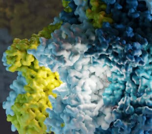
What is cryo-EM?
Cryo-EM: cryo electron microscopy Cryo-electron microscopy (...
Read More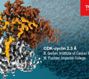
Glacios 2 Cryo-TEM and the Evolution of 200 kV Cryo-EM
Glacios 2 Cryo-TEM and the Evolution of 200 kV Cryo-EM Cryo-...
Read More
Leave a Reply