Cancer organoids are 3D cell cultures derived from patient tumor samples. Cancer organoid models (also known as tumoroid models) can accurately mimic the mutational profiles, gene expression levels, and physical organization of actual tumors, providing a valuable tool for cancer research. However, this increased biological relevancy comes with some differences in how tumoroids are monitored and handled compared to two-dimensional (2D) cell lines, and they exhibit a range of appearances during three-dimensional (3D) culture.
OncoPro Tumoroid Culture Medium was designed to support the growth of tumoroids derived from various cancer types using an easy-to-formulate culture medium that is compatible with both embedded culture within basement membrane extract (BME) hydrogels and suspension culture in non-tissue culture treated dishes and flasks. The use of a standardized culture medium for tumoroid growth removes variability in the cell culture workflow and is best paired with cell culture protocols tailored to tumoroids. This blog post offers practical tips to help you navigate the complexities of cancer organoid culture in the lab and make physiologically-relevant, patient-derived cancer cell culture more accessible than ever.
The Morphology of 3D Cancer Organoids
Tumoroid morphology can differ widely from donor to donor, even within the same type of cancer. This variability underscores the importance of personalized approaches in cancer research and treatment, and of utilizing panels of patient-derived tumoroids that reflect the broad heterogeneity seen in clinical cancer cases. Each organoid model can present donor-dependent growth patterns and structural features. Morphologies can range from cystic structures, where a thin layer of cells surrounds a fluid-filled center, to more solid or grape-like shapes. Representative images of tumoroids over time are shown below to illustrate how tumoroid cells assemble into donor-dependent 3D structures and expand in size over time following the initial seeding of single cells and small cell clusters. During culture, careful monitoring is essential, and we recommend that tumoroids be checked frequently by microscopy. Tumoroids are ready to split when they reach ~100-300 µm in diameter, which occurs within 6-10 days post-seeding for the majority of cultures but may occur after 5-14 days in culture. Overgrowth can lead to necrosis in tumoroid cores, evidenced by the appearance of dark and dense areas during brightfield microscopy. Excessive necrosis may cause cultures to crash, so it is vital to dissociate and reseed tumoroids when they reach ~100-300 µm in diameter for continued expansion. Tumoroids allowed to grow beyond the recommended size range, in particular tumoroid cultures with solid morphology, become difficult to successfully dissociate, inhibiting subsequent cell growth. Frequent imaging of cultures, especially as researchers become accustomed to the morphology and growth rate of patient-derived lines, is vital for success. When best practices for tumoroid feeding and subculture as described below are followed, tumoroid growth rate, genotype, and gene expression can be stable for 250+ days in culture.

Figure 1: Time course images of HuCo021320 colorectal tumoroid growth in OncoPro Tumoroid Culture Medium (suspension culture format, cystic morphology). If tumoroids grow beyond the recommended size range, they develop dark and necrotic regions as seen on Day 9.
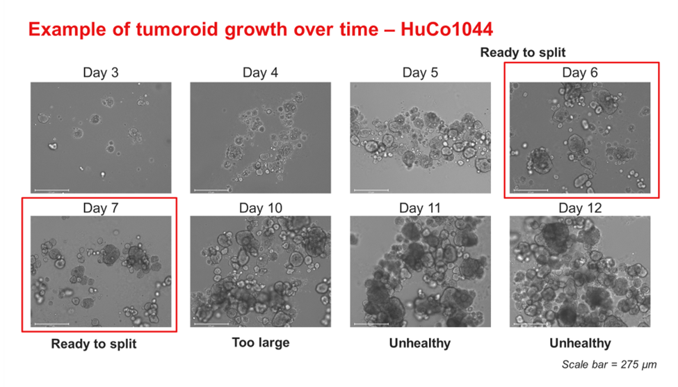
Figure 2: Time course images of HuCo1044 colorectal tumoroid growth in OncoPro Tumoroid Culture Medium (suspension culture format, solid morphology). If tumoroids grow beyond the recommended size range, they develop dark and necrotic regions as seen on Days 11-12.

Figure 3: Time course images of HuLu051421 lung tumoroid growth in OncoPro Tumoroid Culture Medium (suspension culture format, solid morphology). While dark, necrotic cores are not observed in the Day 10 images, tumoroids can become hard to dissociate when the diameter becomes too large and impact the health of subsequent cultures.

Figure 4: Time course images of HuLu051421 lung tumoroid growth in OncoPro Tumoroid Culture Medium (embedded culture format, solid morphology). While dark, necrotic cores are not observed in the Day 10 images, tumoroids can become hard to dissociate when the diameter becomes too large and impact the health of subsequent cultures.
Best Practices for Tumoroid Culture
Tips to help maximize culture success during various stages of the tumoroid workflow are provided below.
Organoid Culture Media Preparation
- Use of OncoPro Tumoroid Culture Medium kit: OncoPro Tumoroid Culture Medium is optimized for the expansion of patient-derived tumoroids. The OncoPro kit streamlines media preparation for cancer organoid culture by eliminating the need to reconstitute, aliquot, and combine individual growth factors and supplements commonly found in homebrew organoid media formulations.

Figure 5: Materials required to prepare OncoPro Tumoroid Culture Medium (excluding antibiotics) and a representative homebrew medium. For homebrew medium, materials required to resuspend, aliquot, and store individual growth factors and supplements are shown.
- Storage of media components: If desired, the components of OncoPro Tumoroid Culture Medium can be aliquoted when volumes of less than 500 ml at a time are needed. OncoPro Basal Medium and OncoPro BSA are stored at 4°C, and aliquots can be taken directly from the original component bottles. The B-27 supplement can be thawed at room temperature, aliquoted, and re-frozen and stored at -20° The OncoPro Supplement should be thawed in a 37°C water bath, aliquoted, flash frozen at -80°C overnight, and then stored at -20°C. Aliquots can be stored up to the expiration date of individual media components. We do not recommend preparing and then freezing complete medium.
- Frequency of media preparation: Prepare complete medium and use within 7 days for optimal performance. Store medium at 4°C when not in use and protect from light.
- Warming media for use: Avoid the use of a 37°C water bath. To warm prepared media for use, we recommend placing the media at room temperature on the benchtop or in a biosafety cabinet for approximately 1 hour, protecting from light. When not in use, store medium at 4°
Cancer Cell Seeding
Dissociated tumoroid cells can be seeded in embedded culture within hydrogels (for example, Geltrex) or in suspension culture, where diluted basement membrane extract (for example, 2% Geltrex by volume) is added to the media to provide extracellular matrix cues.
- Suspension vs. embedded culture: Culture in embedded conditions is often preferred during initial tumoroid derivation (see our derivation application note for more information), while suspension culture facilitates scale up for experiments. Cultures in suspension may need to be split more frequently, as aggregation of free-floating tumoroids can lead to formation of larger tumoroids more quickly than when performing embedded culture (see comparison of HuLu051421 tumoroid line in suspension and embedded culture above in Figures 3 and 4).

Figure 6: Images of HuCo1044 colorectal tumoroids cultured in OncoPro Tumoroid Culture Medium (suspension culture format) at various magnifications on day 7 post-seeding. In suspension culture, tumoroids form 3D cell structures while they are free-floating in medium.
- Addition of BME: Utilize 2% (v/v) solubilized BME in suspension culture to maintain physiologically relevant cell-ECM interactions. These interactions are vital for maintaining the apical-basal polarity of the tumoroids. For best results, drip cold BME into the media after cells and media are added to the culture vessel.
- Use of ROCK inhibitor: Supplement culture medium with 10 µM Y-27632 during initial seeding to support the health of singularized cells. We typically include this compound throughout tumoroid culture, though it could be excluded when medium is exchanged during culture feeding that occurs 2-3 days post-seeding.
Feeding Tumoroids
Tumoroids should be fed regularly to remove metabolic waste and provide nutrients for growth. For embedded cultures, feeding is performed by removing spent media and overlaying fresh OncoPro Tumoroid Culture Medium. For suspension cultures, cells and media are removed from plates or flasks and centrifuged. The supernatant is discarded, and tumoroids are resuspended in fresh medium.
- Feeding frequency: Cells should be fed every 2-3 days using a complete media change to help ensure optimal growth. If not changed every 2-3 days, metabolic waste buildup can negatively impact cell viability and potentially cause the culture to crash. We recommend freshly supplementing media with 10 µM Y-27632 on the day of feeding or subculturing.
- Monitoring medium color: OncoPro Tumoroid Culture Medium, with its slightly acidic pH, may appear more yellow than other culture media. As tumoroids expand and metabolize the medium components, the medium further acidifies, and media may look especially yellow as cells approach passaging. Frequent monitoring (every 2-3 days) is necessary to determine when to split the cultures. If the medium has been changed regularly and tumoroid diameter is within the 100-300 µm range, the yellow color is not a cause for concern.

Figure 7: Examples of media color in tumoroid cultures. Pictured are three T25 flasks imaged 2-3 days post-feeding, all of which contain properly expanding tumoroid cultures. As tumoroid number and size increase and tumoroids approach passaging, media may appear more yellow.
- BME add back: In suspension culture, some of the BME is lost during tumoroid feeding. To replace this lost BME and maintain the benefits of its presence, add 1% (v/v) of BME (such as Geltrex) to the media during each feed. BME should be added after cells and media are plated in the culture vessel instead of being pre-mixed with cells in conical tubes used during feeding.
Passaging Cancer Organoids
Given the variability in morphology from donor to donor, it is essential to monitor tumoroid cultures closely, especially during the initial stages of working with them. As different tumoroids have distinct morphologies and expand at different rates, monitoring helps maximizes the probability of success and provides crucial information about when to subculture the tumoroids.
- Optimal diameter for passaging: We recommend passaging when the diameter of large tumoroids in the culture is 100–300 µm (typically every 6-10 days, though some cultures may be split in the 5–14 day window). Most tumoroid lines reach diameters of 200-300 µm when ready for subculturing, though some lines may only grow to ~100 µm in diameter. Passaging too early can negatively impact viability and lead to excessive dissociation into single cells, which increases the risk of anoikis. Delaying passaging can result in necrotic core formation, ineffective dissociation, metabolic waste buildup, and eventual culture crash. The tumoroids in the image below are representative of the variations in morphology that can occur within a given cancer indication, in this case colorectal cancer.

Figure 8: Representative images of three colorectal cancer tumoroids lines that are ready to passage at 10x, 4x, and 1.25x magnification. Scale is indicated.
- Washing protocol: Use cold buffers (e.g., cold DMEM/F12, DPBS -/-) to break up the polymerized BME and separate cells from the BME network. This step is critical to ensure that enzymes in dissociation reagents effectively dissociate the tumoroids.
- Dissociation reagents: In our experience, using StemPro Accutase for splitting has yielded steady growth rates. This enzyme efficiently dissociates tumoroids into single cells and small cell clusters without compromising cell viability. StemPro Accutase aliquots should be stored at -20°C and thawed at room temperature on the day of use. Other dissociation reagents such as TrypLE could potentially be used, depending on the specific requirements of the study and the characteristics of the organoid model. We recommend supplementing the dissociation reagent with 10 µM Y-27632 during incubation to support cell health of singularized tumoroid cells.
- Dissociation temperature: We recommend dissociating tumoroids at 37°C in StemPro Accutase supplemented with 10 µM Y-27632. This can be performed in a 37°C water bath or on a CO2 resistant shaker placed inside a 37°C, 5% CO2 cell culture incubator and set at 120 rpm. Do not use a heated bead bath for dissociation. For dissociation in a water bath, gently swirl the conical tube containing the tumoroid suspension every 2-3 minutes to maintain even dispersion within the dissociation reagent.
- Trituration: After incubation with the dissociation reagent, triturate the cell suspension 20 times with a P1000 pipette tip to further break up tumoroids. Do not use wide bore pipet tips, as these will not provide sufficient mechanical force to disrupt the 3D structures. The pipette tip should be pre-wetted (via trituration of cell-free OncoPro Tumoroid Culture Medium) prior to this step to avoid adhesion of cells to the pipette tip. Adequate dissociation is indicated when the cell solution turns cloudy after trituration with a P1000 pipette tip. There should be no clusters visible by the naked eye, and no intact tumoroids with defined borders should remain immediately post-passage.

Figure 9: Images of conical tubes containing effectively dissociated tumoroids (top) and tumoroids needing additional incubation time in StemPro Accutase (bottom), which are visible by eye.
- Extended incubation: If in doubt, incubate in the dissociation reagent for an additional 5-10 minutes and triturate again before diluting and counting. We recommend increasing the dissociation time in two minute increments, up to 20 minutes total, until cells are properly dissociated.
- Vortexing difficult lines: For lines that are particularly challenging to dissociate effectively, consider briefly vortexing (e.g., 2s using using FisherbrandTM Mini Vortex Mixer) the closed tube every two minutes during incubation with the dissociation reagent.
- Cell counting: We recommend counting cells at each passage to better control the consistency of seeding and quantify the doubling times and cumulative population doublings of each model. During counting, small clusters of cells may be present. Individual cells present in these clusters should be counted to obtain accurate data (see Figure 10). Decluster algorithms can be implemented on image-based automated cell counters to accurately count cells present in small clusters. We have found that setting minimum object diameter to 8 µm and maximum object diameter to 60 µm and using the highest degree of declustering possible on image-based automated cell counters generates accurate cell count data.
- Cell viability during subculturing: Tumoroid cell viabilities are variable and can differ substantially from donor to donor. After dissociation, tumoroid viability (by Trypan Blue exclusion) is typically between 65-90%. While these viabilities are lower than those typically seen with immortalized cell lines, they should remain stable over multiple passages and do not necessarily indicate poor cell health in the culture.
After dissociation, cultures should consist of a mixture of single cells and small cell clusters, as shown below.

Figure 10: Representative images of tumoroids post-dissociation, with live (green) and dead (red) cells indicated. When counting, attempt to count individual cells that are present in clusters for the most accurate quantification of cell number.
Other Considerations for Cancer Organoid Culture
- Cell adhesion during suspension culture: Some tumoroids are innately “sticky” and may adhere to non-tissue cultured treated flasks during suspension culture. If cell adhesion is observed for a particular tumoroid line, coat well plates or flasks with 7.5% BSA solution (diluted in DPBS without calcium or magnesium) for 30 minutes in a 37°C cell culture incubator prior to seeding cells. The BSA solution can be aspirated immediately prior to adding tumoroid cell suspensions in OncoPro Tumoroid Culture Medium at initial seeding or post-feed, and no intermediate wash steps are required.
- Cell adhesion during embedded culture: In some cases, cell adhesion and spreading may be observed during culture within BME hydrogels. To help minimize cell adhesion to cell culture plastics, non-tissue culture treated well plates and dishes can be used. If handled carefully, hydrogel domes will remain in place on these vessels when they are inverted to polymerize BME during the initial cell seeding protocol. If desired, adherent cells can be detached using TrypLE or similar dissociation reagents after tumoroid cells in BME domes are collected.
- Pre-wetting of serological pipettes and pipette tips: We recommend pre-wetting serological pipettes and pipette tips prior to tumoroid handling to minimize cell/tumoroid adhesion to culture plastics and subsequent loss of cells. Pre-wetting may not be required for workflows that tolerate loss of cells during handling.
Advancing Cancer Research with OncoPro Tumoroid Culture Medium
Cancer organoid models grown in offer a powerful platform for studying tumor biology. By understanding and leveraging cancer organoids, researchers can advance the field of cancer research and contribute to the development of more effective treatments. Regular imaging, consistent feeding, and appropriate subculturing are key practices that help ensure the viability and robustness of these 3D models. Detailed protocols and recommended cell densities and media volumes are included in the OncoPro Tumoroid Culture Medium User Guide.
Note: Some images adapted from Paul, C.D. et al. bioRxiv 10.1101/2024.06.10.598331v1 (2024).
Related Content
Cancer organoids vs cancer spheroids
For Research Use Only. Not for use in diagnostic procedures.