What type of T cell stimulation protocol should I use for my experiments?
Stimulation of peripheral blood mononuclear cells (PBMCs) is useful to expand and differentiate populations of immune cells. Often, stimulated cells will express different cell surface receptors, up or down regulate expression of intracellular cytokines, and modify proteins, as compared to resting or naïve cells. Stimulants can include phorbol 12-myristate 13-acetate (PMA), lipopolysaccharide (LPS), staphylococcal enterotoxin B (SEB), antibodies, or peptide pools [2]. The appropriate stimulant and stimulation condition are dependent on the cell type and cytokine of interest.
CD3 zeta phosphorylation is an indicator of T cell activation via the TCR complex, and treatment of T cells with monoclonal antibodies that engage the TCR can be used as a model for antigen-induced activation [3,4]. The first step in this process is to treat cells with anti-CD3 antibodies to engage the TCR, and anti-CD28 antibodies to provide a co-stimulatory signal. This is followed by a secondary antibody that recognizes the host IgG of the anti-CD3 and -28 antibodies, to cross-link the subunits of the TCR complex for optimal signal transduction and T cell activation.
How do you know if my T cell stimulation protocol worked?
TCR activation involves the cytoplasmic tails of the CD3 subunits: CD3 gamma, CD3 delta, CD3 epsilon and CD3 zeta (alternatively named CD247). One of the early steps in the TCR cascade include CD3 zeta phosphorylation. It occurs immediately downstream of Lck activation, and upstream of ZAP-70, LAT, and SLP-76, and allows for direct identification of TCR engagement. While Lck activation is upstream of CD3 zeta phosphorylation, it is also involved in other receptor signaling pathways, such as IL-2 and CD2, making CD3 zeta a more TCR-specific method of identifying T cell activation.
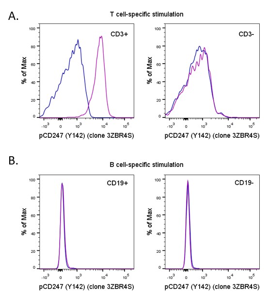
Based on known expression patterns, phospho-CD247 (Tyr142) stains stimulated CD3+ T cells following a T cell-specific stimulation with no staining observed without stimulation (figure 1A), in CD3- cells, or in cells stimulated with a B cell-specific stimulation (figure 1B).
Figure 1: Intracellular staining of stimulated human peripheral blood cells. (A) Human peripheral blood cells unstimulated (blue histogram) or stimulated for 15 minutes with CD3 (clone OKT3), Functional Grade and CD28 (clone 28.6), Functional Grade, followed by a 5 minute incubation with F(ab’)2-Goat anti-Mouse IgG (H+L) Secondary Antibody, Functional Grade (purple histogram) were fixed and permeabilized with the IC Fixation & Permeabilization Buffer Set and protocol, followed by intracellular staining with CD3 (clone UCHT1) and Phospho-CD247 (Tyr142) (clone 3ZBR4S). Staining of Phospho-CD247 in CD3+ cells is shown on the left, and on CD3- cells on the right, with no stimulation observed in CD3- cells. Cells in the lymphocyte gate were used for analysis. (B) Human peripheral blood cells unstimulated (blue histogram) or stimulated for 5 minutes with F(ab’)2-Goat anti-Human IgG, IgM (H+L) Secondary Antibody, Functional Grade (purple histogram) were fixed and permeabilized with the IC Fixation & Permeabilization Buffer Set and protocol, followed by intracellular staining with CD19 (clone HIB19) and Phospho-CD247 (Tyr142) (clone 3ZBR4S). Staining of Phospho-CD247 in CD19+ cells is shown on the left, and on CD19- cells on the right, and no stimulation is observed in either group following this B cell-specific treatment. Cells in the lymphocyte gate were used for analysis.
What type of research can utilize phospho-CD247?
Those using the phospho-CD247 antibody would likely be studying T cell activation and CAR T cells. The zeta chain of the CD3 complex is the part responsible for initiating the signaling cascade upon engagement of the T cell receptor. It is phosphorylated on Tyr142 upon TCR activation, and functions to recruit ZAP70, which further propagates the signal. Staining for phospho-CD247 would demonstrate TCR receptor activation and functionality. In particular, this antibody would be useful for the CAR T workflow, as this would be a way to determine that the engineered TCR is functional and can receive and propagate a signal.
If incorporating CD247 into a flow cytometry panel, what other markers would be recommended for use and why?
As far as other markers that would be associated with it, some of the classic T cell markers, such as CD3, CD8, and CD28 would be appropriate. We also have antibodies against other phosphorylated proteins involved in T cell signaling, such as phospho-ZAP70, phospho-LCK, and phospho-SLP76.
What can you tell me about the relative expression of CD247 and picking a fluorophore conjugate?
CD3 is highly expressed and can be stained with both brighter fluorophores such as PE, APC, and PerCP-eFluor 710 conjugates, but also dimmer ones too including FITC or eFluor 450.
Where can I find information about the key antibodies to T cell markers that are available?
The first resource we recommend is the Thermo Fisher Scientific website. All listed antibodies are presented with a description of the antigen they recognize, and the flow plot data on primary human or mouse cells. Application notes are also available with detail protocols and guidance with gating. For building a T cell panel we recommend reading our application notes including a 10-color T cell panel. Then, try building your own immunophenotyping panel with a comprehensive panel builder.
Key T cell References:
- Lin J, Weiss A. (2001) T cell receptor signaling. J Cell Sci. 114: 243-4.
- Kay, J.E. (1991). Mechanisms of T lymphocyte activation. Immunology Letters. 29, 51 – 54.
- Trickett A, Kwan YL. (2003) T cell stimulation and expansion using anti-CD3/CD28 beads. J Immunol Methods. 275(1-2):251-5.
- Risueño RM, Schamel WW, Alarcón B. (2002) T cell receptor engagement triggers its CD3epsilon and CD3zeta subunits to adopt a compact, locked conformation. PLoS One. 3(3):e1747.
For Research Use Only. Not for Use in Diagnostic Procedures.
_______________________________________________________________________________________________________________________________________________________________________________________

Natalie Oxford, antibody development scientist at Thermo Fisher Scientific, is responsible for the development of anti-phospho CD247 by flow cytometry. She has over 11 years of R&D experience and is providing her guidance in adding antibodies like anti-phospho CD247 to complex immunophenotyping flow cytometry applications.
hi Natalie,
I have a quick question about this TCR zeta phosphorylation.
I am stimulating mouse T cells, for 3 days, in vitro with anti-CD3 . My question is “would I able to detect Phospho-CD247 even after 3 days of stimulation? Please let me know, Thanks a lot