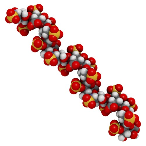
 Researchers studying deceased patients or rare diseases have a unique problem in that there may be only a finite number of banked samples available to study. For this reason, getting quality samples is extremely important. Since additives in the blood—particularly heparin—can interfere with downstream analysis, manufacturers of enzyme-linked immunosorbent assays (ELISA) warn that samples containing more than 3U/μL of heparin are not usable for testing. Moreover, the negatively charged glycosaminoglycans (GAG) derivatives in heparin can also interfere with RT-PCR, microarray or RNASeq profiling assays.
Seeking a solution to this incompatibility, researchers Sánchez-Fito and Oltra discovered a way to make heparinized blood samples suitable for ELISA and RT-PCR amplifications.1 The team developed a low-cost and effective protocol based on earlier work from Plieskatt et al.2 In the earlier study, the authors treated blood samples with Bacteroides Heparinase I to break down heparin. They found it significantly improved RTqPCR data.
For their experiments, Sánchez-Fito and Oltra et al. used Bacteroides Heparinase I, Heparinase II and Heparinase III at concentrations of 0.05–0.1 U/μL to break down heparin PBS, heparinized plasma or heparin spiked preparations. Using a colorimetric reporter assay with toluidine blue solutions as a reporter, the team employed a NanoDrop 2000 UV-VIS spectrophotometer (Thermo Scientific) to measure the absorbance between 400 nm and 750 nm of 0.005% toluidine blue solutions for heparin at 1.7–17 IU/mL. They identified the most significant wavelengths for the discrimination of solutions containing levels above or under 3 U/μL.
The team noted that solutions containing 1.7 IU/mL of heparin present clear differences in absorbance both at 510 nm and 630 nm when compared with those containing 3.4 IU/mL of heparin. However, in PBS or plasma below 1.7 IU/mL, they did not see a significant change in absorbance at the optimized wavelengths, indicating heparin could not be detected at concentrations of 0.85 IU/mL or below.
The researchers maintain this work demonstrates a cost-effective method to make heparinized, otherwise unusable, blood-derived collections suitable for analysis by ELISA and RT-PCR amplifications. This, in turn, will help enhance research opportunities for valuable samples.
References
1. Sánchez-Fito, M.T. and Oltra, E. (2015) “Optimized treatment of heparinized blood fractions to make them suitable for analysis,” Biopreservation and Biobanking, 13(4) (pp. 287–95), doi: 10.1089/bio.2015.0026.
2. Plieskatt, J.L., et al. (2015) “Circumventing qPCR inhibition to amplify miRNAs in plasma,” Biomarker Research, 2(13), doi: 10.1186/2050-7771-2-13.
Leave a Reply