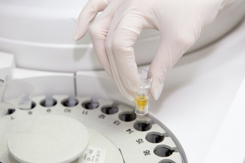
 Urine sample preparation can greatly impact downstream experiments. For example, measuring creatinine and cystatin C levels gives both important insights into the total protein concentration and clues as to how well the body’s filtration systems are functioning, but if the samples are not prepared properly, the resulting measurements may be altered. In a recent issue of Biopreservation and Biobanking, Ammerlaan et al. evaluated urine preparation techniques.1 This group of investigators provided a validated methodology for preparing cell-free and debris-free urine supernatant samples.
The team collected 100 mL urine samples from one male and one female who were both healthy at the time of the study. They spun the samples in either a 5810R Eppendorf centrifuge or a Thermo Scientific SL40 FR centrifuge. They evaluated three speeds: 1,000 g and 2,000 g (both for 10 minutes with a medium brake) and 10,000 g (20 minutes with a hard brake), all at room temperature (19°C–24°C). They also evaluated double centrifugation by transferring 13 mL to 15 mL high-performance conical tubes at 2,000 g for 10 minutes with a medium brake, then 12,000 g for 10 minutes with a hard brake.
Taking into account the sample storage temperature, they further assessed whether refrigerated temperatures would improve samples by minimizing degradation and possible microbial growth. To do this, they spun samples at 4°C at 12,000 g for 20 minutes with a medium brake. They compared sample robustness/brake speeds at 4°C at 12,000 g for 20 minutes, with soft, medium and hard brakes. To compare robustness/temperature, they ran samples at 4°C versus 21°C, at 12,000 g for 20 minutes, with a hard brake. To assess the reproducibility, they split one urine sample into three aliquots and ran them at 12,000 g, at 4°C for 20 minutes, with a hard brake.
They also evaluated the reproducibility by using microparticle counts as well as the cystatin C and creatinine concentrations as acceptance criteria. They used the Creatinine Parameter Assay Kit to measure creatinine levels and the Human Cystatin C Quantikine ELISA Kit to measure cystatin C. To provide further validation, the team also used a metabolomics workflow by performing gas chromatography and mass spectrometry.
The initial analysis revealed that the higher centrifugation speeds resulted in ideal lower microparticle counts. The team remarked that the two-step centrifugation is optimal for intact cell isolation; however, if intact cells are not an imperative, a single high-speed centrifugation results in lower microparticle counts and is less time consuming, with a reduced chance of handling errors.
Ultimately, the investigators came to the conclusion that the optimal protocol for preparing urine samples is a 20-minute, 12,000 g centrifugation at 4°C with a hard brake. These conditions also resulted in superb reproducibility in terms of mean microparticle counts. Additionally, the cystatin C and creatinine concentrations were within acceptable limits.
The authors present this work to improve sample handling for biospecimens in biobanks and other clinical laboratories while also acknowledging the limitations of the small number of samples used in this study. Because of this, they advise that negative results should be interpreted with caution.
Reference
1. Ammerlaan, W., et al. (2014) “Method validation for preparing urine samples for downstream proteomic and metabolomic applications,” Biopreservation and Biobanking, 12(5) (pp. 351–357), doi:10.1089/bio.2014.0013.
Leave a Reply