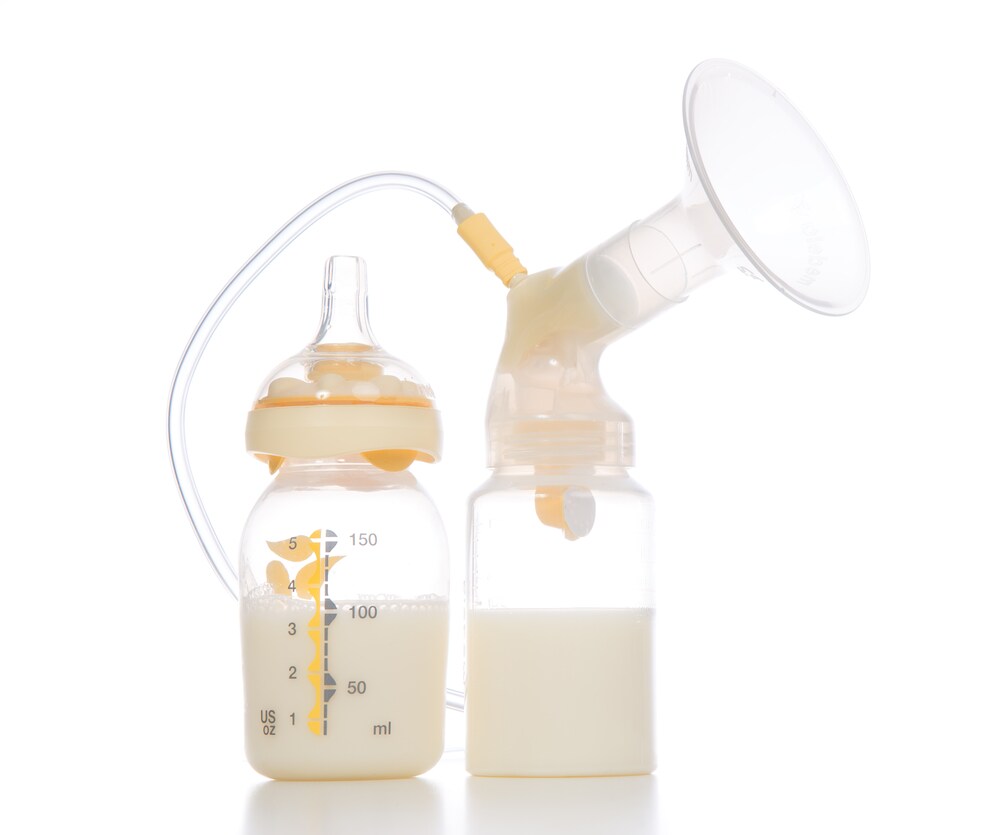
 Human milk sufficiently meets all growth and developmental needs for infants by way of a unique nutrient composition, antibodies, and metabolic enzymes. While several other research groups have studied the components of human milk, according to Dall’Asta et al., these earlier studies fail to address the differences in an infant’s digestion systems compared with that of adults.1
Dall’Asta and colleagues developed a unique model to simulate a newborn baby’s digestive system. They based their strategy on a 10-year-old study that used a more general in vitro digestive model.2 After applying the newly established infant digestive model to human milk samples, Dall’Asta and colleagues analyzed digested peptides using micro liquid chromatography mass spectrometry (µLC-MS).
Six samples of human breast milk were obtained following strict criteria. All women were Caucasian between 25 and 45 years old. They had all been lactating for 40 days (±2 days) calculated from the date of delivery and were all on their second pregnancy which lasted for >37 weeks. They had no pre-eclampsia with normal weight (body mass index 418.5 and 525 kgm2) and no history of smoking.
The team subjected milk samples to the model digestive system which simulated three normal digestive stages: salivary, gastric, and intestinal. To represent the salivary phase, the team added lyophilized α-amylase to milk samples. For the gastric phase, the team used 2.6 ml of a mix of gastric juices previously developed. They also corrected the pH to 4.5 with 1 M HCl. Next, they mixed samples with a pepsin and NaCl solution and stirred at 37℃ for 35 min. They added duodenal juices and bile salts, and then stirred the sample before adding a β-galactosidase, a key enzyme in the infant digestive process.
To further simulate intestinal conditions, they stirred the mixture and then transferred it to a 15 cm dialysis tube with glass marbles to favor the mixing. Then they transferred the dialysis tube to an airtight container containing gastric juice and bile salts and incubated it at 37℃ for 2 h.
After each digestive phase the team removed small aliquots for analysis. They performed sodium dodecyl sulfate polyacrylamide gel electrophoresis (SDS-PAGE) to analyze α–lactalbumin (α-LA), the major protein in breast milk. They found α-LA remained undigested during the salivary and gastric phases, but it disappeared completely after intestinal digestion.
For µLC-LTQ-MS, the researchers used an LTQ Orbitrap mass spectrometer (Thermo Scientific) and Proteome Discoverer (Thermo Scientific) for the data analysis.
The team identified 149 peptides from β-casein, 30 peptides from α-LA, 26 peptides from αs1-casein, 24 peptides from k-casein, 28 peptides from osteopontin, and 29 from lactoferrin. They also identified hydrolyzed caseins, a-lactalbumin, and osteopontin, which was not previously reported in earlier studies. The authors maintain this work is an good starting point to more fully understand the properties of human milk.
Learn more about food analysis in our Food and Beverage Community
References
1. Dall’Asta, C. et al. (2015) “Development of an in vitro digestive model for studying the peptide profile of breast milk.” International Journal of Food Sciences and Nutrician, 66:4, (pp. 409-415)
2. Versantvoort CHM, (2005). “Applicability of an in vitro digestion model in assessing the bioaccessibility of mycotoxins from food.” Food Chemistry and Toxicology 43: (pp. 31–40)


Leave a Reply