Electron microscopy (EM) allows us to observe a world exponentially smaller than the one we can see with our unaided eyes or even with the familiar light microscope. Electron microscopy uses electrons to “see” small objects in the same way that light beams let us observe our surroundings or objects in a light microscope. With EM, we can look at the feather-like scales of an insect, the internal structures of a cell, individual proteins or even individual atoms in a metal alloy.
TEM vs SEM Comparison
The two most common types of electron microscopes are transmission (TEM) and scanning (SEM) systems, but the differences between these two instruments can be fairly nuanced. Here we hope to provide a fundamental primer for individuals looking to get started with this powerful technique.
Scanning Electron Microscope (SEM)
Imagine you are in a dark room with a weak flashlight. To explore your surroundings, you might sweep the light across the room, much like someone reading a book: left to right and top to bottom. SEM functions similarly, sweeping the electron beam across the sample and recording the electrons that bounce back. This technique allows you to see the surface of just about any sample, from industrial metals to geological samples to biological specimens like spores, insects, and cells. While SEM cannot see features to the same level of detail as TEM, it is much faster, less restrictive, and can sometimes be performed with limited or no sample preparation.
Transmission Electron Microscope (TEM)
When a movie plays in the theater, light is transmitted through an image on a film. As the beam of light passes through, it is modified by the image and the contents of the film are then displayed. TEM works in the same way but with electrons, passing through, or transmitting, an ultrathin sample to a detector below. TEM allows you to observe details as small as individual atoms, giving unprecedented levels of structural information at the highest possible resolution. As it goes through objects it can also give you information about internal structures, which SEM cannot provide. TEM is, however, limited to samples that can be thin enough to let electrons pass through them. This thinning process is technically challenging and requires additional tools to perform.

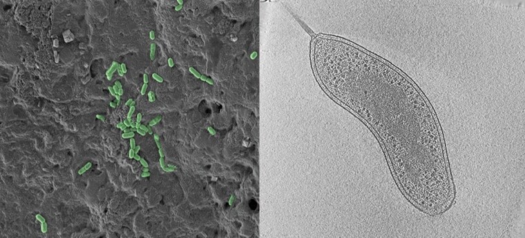
SEM (left) and TEM (right) images of bacteria. Whereas SEM shows numerous bacteria on a surface (green), the TEM image shows the interior structure of a single bacterium.
Overall, TEM offers unparalleled detail but can only be used on a limited range of specimens and tends to be more demanding than SEM. It is important to note that advanced techniques such as cryo-EM, a method which looks at typically biological specimen in a vitrified, amorphous state, have expanded the capabilities of TEM significantly. In particular, biomedical and pharmaceutical research may benefit from the molecular and cellular details that can be revealed by cryo-EM.
In general, if you need to look at a relatively large area and only need surface details, SEM is ideal. If you need internal details of small samples at near-atomic resolution, TEM will be necessary.
Learn more about electron microscopy >>
//
Alex Ilitchev, PhD, is a Scientific Content Writer at Thermo Fisher Scientific.

Leave a Reply