Cryo-electron microscopy (cryo-EM) is becoming an indispensable tool in pharmaceutical research due to its ability to provide high-resolution molecular insights into infectious, neurological, and hereditary disorders, complementing existing tools for structure-based drug design. We recently hosted a virtual symposium to showcase how advances in cryo-EM have enabled structure-based insights for the drug discovery pipeline.
Experts from the biopharmaceutical industry and academia shared their recent successes along with practical considerations that guide their cryo-EM work. The presentations and discussions, highlighted below, are now available online in the Drug Discovery Virtual Events section of our Media Gallery.
Latest scientific developments in cryo-electron microscopy and electron diffraction: two keynotes
Electron diffraction has a lot in common with X-ray crystallography and acts as a complementary method, generating structural information from smaller crystals that are unsuitable for X-ray analysis.
As part of the “Latest Scientific Developments” keynote session of the symposium, Professor Paul Midgley of Cambridge University discussed the development and applications of electron diffraction, including “continuous rotational electron diffraction,” popularly known as MicroED. A particular focus was placed on the development of scanning electron diffraction (SED) and its use in microstructure determination for pharmaceuticals.
Cryo-EM in drug discovery at AstraZeneca
In the second half of the keynote session, Dr. Christopher Phillips of Astra Zeneca discussed how his team supports pipeline projects with cryo-EM insights. Notably, Dr. Phillips highlights the significance of cryo-EM in the study of membrane protein and large macromolecules that have historically been challenging for X-ray crystallography. Dr. Phillips also talked about the Cambridge Pharmaceutical Cryo-EM Consortium, a collaboration between Medical Research Council Laboratory of Molecular Biology, University of Cambridge, Thermo Fisher and five pharmaceutical companies, to enable shared training and access to cryo-EM instrumentation.
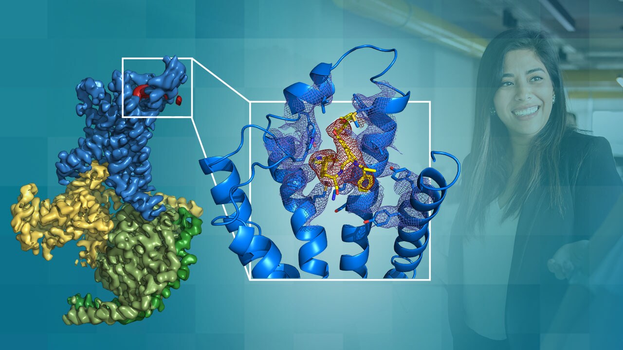
Cryo-EM solution optimized for drug discovery: structure-based design and GPCR research
G-protein-coupled receptors (GPCRs) are a large class of membrane proteins that have been implicated in an extraordinary variety of diseases including diabetes, heart disease, Alzheimer’s, and many more. It is estimated that over one-third of current marketed drugs act on GPCRs. As membrane proteins, they have been difficult to study with traditional methods and are a prime target for cryo-EM structural analysis.
Professor Patrick Sexton and his group at Monash University are leaders in GPCR research and are actively utilizing cryo-EM as part of their work. In his talk, Prof. Sexton discussed the critical aspects of structure-based discovery: throughput, robustness, and resolution, and how cryo-EM has met these factors. Additionally, the group’s recent work on the glucagon-like peptide-1 receptor (GLP-1R) was highlighted.
Cryo-EM workflow round table discussion
Industry experts talked about their experience integrating cryo-EM into their drug discovery workflow and bringing cryo-EM instrumentation in-house. The panel focused on covering the accessibility of the technique and the efforts being made to increase the throughput, accuracy, and scope of cryo-EM for structure-based pharmaceutical research.
Visit our Cryo-EM in the Pharmaceutical Industry webpage to learn more about how pharmaceutical companies are using cryo-EM to accelerate their drug discovery.
To find out how you can incorporate cryo-EM into your pharmaceutical research, please see our Drug Discovery Using Electron Microscopy page.
Alex Ilitchev, PhD, is a Science Writer at Thermo Fisher Scientific.
//
To learn more about electron microscopy for Life Sciences, please visit our Learning Center.
Subscribe Now to receive new Accelerating Microscopy posts straight to your inbox.
Guiding the Development of an HIV Vaccine with Structure-Based Drug Design
Novel drug development in the fight against the HIV pandemic...
Read More
Virus microscopy unravels chikungunya virus structure
Global impacts of the chikungunya virus Chikungunya is a vir...
Read More
What is cryo-EM?
Cryo-EM: cryo electron microscopy Cryo-electron microscopy (...
Read More
Glacios 2 Cryo-TEM and the Evolution of 200 kV Cryo-EM
Glacios 2 Cryo-TEM and the Evolution of 200 kV Cryo-EM Cryo-...
Read More
Leave a Reply