 A wealth of valuable proteomics data is available in tissue archives. However, since most of this library is stored as formalin-fixed, paraffin-embedded (FFPE) tissue blocks, its value to proteomic discovery has been somewhat limited so far. In order to maximize use of this resource, Drummond et al. (2015) recently demonstrated a workflow that prepares protein extracts from specific cell types for onward mass spectrometry–based proteomics analysis.1 Using archival FFPE brain tissue blocks, the team validated a method that enabled proteomic characterization and quantitation using neurons from patients with severe Alzheimer’s disease (AD).
Currently, proteomics researchers focus more on using snap-frozen tissue samples, since the workflow is established and validated. However, studies require large amounts of tissue for preparation, and it is difficult to focus in on a specific cell type within the sample. Drummond et al. used laser capture microdissection (LCM) to select neurons in 10-year-old FFPE tissue blocks of temporal cortex collected from two patients with advanced AD prior to sample preparation for liquid chromatography–tandem mass spectrometry (LC-MS/MS) proteomic characterization.
The researchers investigated a number of variables in the new workflow, looking at tissue volume, efficiency of LCM to isolate neuronal material, tissue digestion and protein solubilization methods required specifically for AD pathology. Following tissue deparaffinization, protein extraction and digestion, the team desalted the preparations using POROS beads before proteomic analysis using an EASY-nLC 1000 high-performance LC system coupled with a Q Exactive mass spectrometer (both Thermo Scientific). Drummond et al. searched for peptide matches in the UniProt human database using the SEQUEST tool within Proteome Discoverer software (Thermo Scientific).
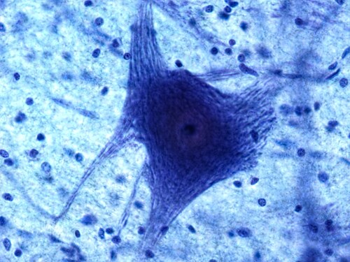
Using cresyl violet staining, the scientists showed that LCM efficiently captured only the neurons required for the study, thus excluding the surrounding tissues from further analysis. They also demonstrated the β amyloid accumulation that is diagnostic for AD in the tissue sections under investigation using specific immunohistochemistry (IHC). The team also found that using the stain or IHC confirmation did not affect subsequent proteomics results; prior treatment did not cause protein loss or disrupt the cells.
The research team found that all sample preparation methods worked well, with no obvious advantage gained from using detergent in the lysis buffer. By omitting the detergent in the buffers, the team prevented excess tissue loss during processing—a valuable step in preserving limited samples. However, they did find that adding formic acid extraction (FAE) to the lysis reagents enhanced the final proteomic analysis. This was due to increasing availability of insoluble protein aggregates and low-abundance proteins such as D-tau. Although FAE reduced the number of peptides per protein, it did increase the overall totals identified.
The researchers also investigated how much tissue was required for satisfactory proteomic analysis to arrive at minimum sample volumes for the workflow. For illustration, a 10 mm2 cortical area yielded approximately 80,000 neurons on initial counting. From this, the team prepared samples ranging from 0.5 mm2 upwards for LC-MS/MS. In terms of unique proteins identified, the team achieved a plateau between 0.5 mm and 3 mm2. A sample size of 2 mm2 (20,000 neurons) gave a total of 956 proteins identified, whereas 0.5 mm2 (4000 neurons) yielded 399.
Referring to protein identification databases in order to validate the suitability of the workflow for concentrating on specific cell types, Drummond et al. found that 77% (306/399) of proteins detected in the 0.5 mm2 samples were neuronal in origin. Furthermore, comparison with previous studies recorded in the UniProt human database showed that 52% (209/399) of the proteins identified within the LCM-LC-MS/MS workflow had already been logged as associating with AD.
Drummond et al. suggest that the validation steps taken using FFPE tissue collected from AD patients show that the proposed LC-MS/MS workflow is suitable for localized proteomics where researchers would like to focus on a specific population of cells. Moreover, the results show that FFPE can be used with confidence for proteomic characterization.
Reference
1. Drummond, E.S. et al. (2015) “Proteomic analysis of neurons micro-dissected from formalin-fixed, paraffin-embedded Alzheimer’s disease brain tissue,” Nature Scientific Reports Scientific Reports, 5(15456). doi: 10.1038/srep15456.
Post Author: Amanda Maxwell. Mixed media artist; blogger and social media communicator; clinical scientist and writer. A digital space explorer, engaging readers by translating complex theories and subjects creatively into everyday language.
Leave a Reply