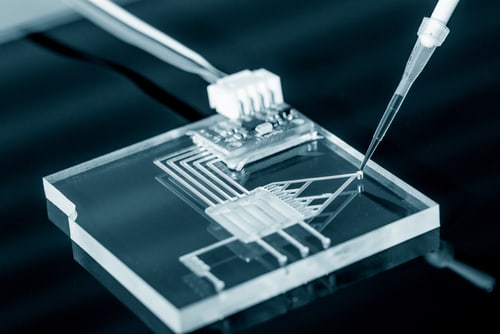
 Microfluidic devices offer novel time-saving advantages to researchers using mass spectrometry (MS) for proteomic analysis, and these tools have been becoming more available in MS over the last two decades. Recent papers by Redman et al. (2016) show how coupling microfluidics capillary electrophoresis with mass spectrometric analysis is a useful tool for characterizing intact monoclonal antibody-drug conjugates (ADCs) and for monitoring glycated hemoglobin in diabetes management.1,2
Despite advantages in reduced sample-handling and preparation times alongside an attendant decrease in error, researchers may not be familiar with the role that microfluidics could play in mass spectrometry-based analytical workflows. They may be more familiar with reports of coupling problems between microfluidics devices and MS instrumentation that hinder efficient use. However, that view is worth revising: a recent announcement from Thermo Fisher Scientific Inc. and a product demonstration at June’s American Society for Mass Spectrometry meeting in San Antonio, Texas, show that the 908 Devices ZipChip is fully compatible with Thermo Scientific Exactive series, Q Exactive series and LTQ Orbitrap Hybrid FT mass spectrometers.
So how can microfluidics aid mass spectrometric detection and analysis? According to Wang et al. (2015), in a review of proteomics papers published between 2010 and 2014, the miniaturization involved in microfluidics is of direct benefit to mass spectrometric analysis.3 Since microfluidic chips are small and compact, and can handle multiple sample processes, they offer benefits in terms of sample volume, reagent use and processing speed.
Microfluidic devices or chips are capable of integrating multiple sample-preparation processes, such as trypsin digestion, solid phase electrophoresis (SPE) and electrophoretic separation, often all within the same assembly. Since the chips handle very low “microscale” volumes, researchers can use less sample in addition to sparing reagents. Furthermore, integrating multiple processes reduces handling errors in the workflow in addition to speeding up preparation: the 908 Devices ZipChip, for example, has a three-minute sample-preparation turnaround.
In their review, Wang et al. present the different ways in which researchers have successfully coupled microfluidic devices, by both integrated and external emitters, to MS instrumentation. Covering the three main forms of microfluidics—analog or conventional channel-based, droplet and digital—and coupling to ionization methods including electrospray; MALDI; and paper-based, plasma and surface acoustic wave nebulization, the review forms a comprehensive catalog of approaches available to researchers. Although scientists experienced problems with altered fluid dynamics from integrated emitters, the numerous papers covered in the review describe how research groups overcame these obstacles to present sample preparations directly to MS instrumentation without additional handling.
As discussed in both papers by Redman et al., the integrated microfluidics platform provides in-line separation by capillary electrophoresis within the chip itself. The device then couples directly with the electrospray ionization (ESI) source and mass spectrometer. Minimal sample preparation enables researchers to characterize intact ADCs by mass shifts and changes in mobility during MS analysis. The second paper describes how the microfluidics platform also achieves in-line separation compatible with ESI-MS, allowing researchers to determine circulating glycosylated HbA1c hemoglobin levels as an indication of glycemic control in diabetes. Furthermore, the platform also enables evaluation of levels of human serum albumin glycation, thus giving a better overview of short- and long-term patient control.
As shown by Wang et al. in their review and in the recent press release, coupling this new microfluidics technology to MS instrumentation is becoming more straightforward. For example, the 908 Devices chip couples directly with certain Thermo Scientific mass spectrometers for an uninterrupted workflow. Taking advantage of what Redman et al. describe as a “simple, generic strategy for characterization with direct mass spectrometric analysis” should become increasingly available to researchers.
References
1. Redman, E.A., et al. (2016) “Characterization of intact antibody drug conjugate variants using microfluidic capillary electrophoresis−mass spectrometry,” Analytical Chemistry, 88(4) (pp. 2220−2226).
2. Redman, E.A., et al. (2016) “Analysis of hemoglobin glycation using microfluidic CE-MS: A rapid, mass spectrometry compatible method for assessing diabetes management,” Analytical Chemistry, 88(10) (pp. 5324−5330).
3. Wang, X., et al. (2015) “Microfluidics-to-mass spectrometry: A review of coupling methods and applications,” Journal of Chromatography A, 1382 (pp. 98–116).
Leave a Reply