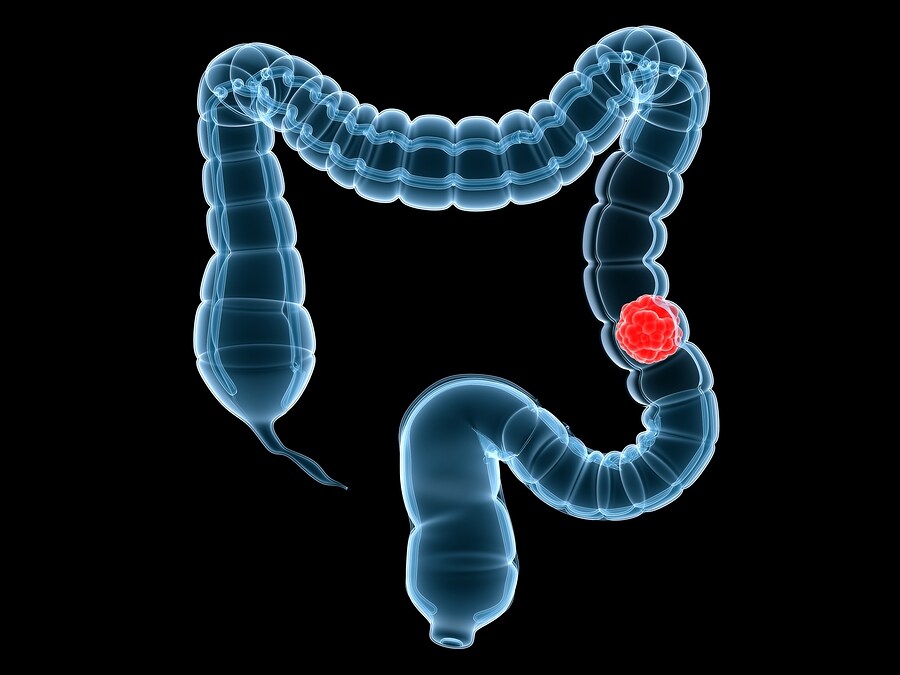
Cancer Tumor in the Colon
Cell lines LIM1215, LIM1899, and LIM2405 were obtained for analysis. Cells were incubated, the RNA was extracted using Trizol, and samples were run in triplicate. RNA was sequenced. For proteomic analysis, proteins were separated using SDS-PAGE gels and digested with trypsin. Liquid chromatography-tandem mass spectrometry (LC-MS/MS) was performed using an LTQ-XL Linear Ion-Trap mass spectrometer (Thermo Scientific) coupled to an in-house packed chromatography column (Agilent Technologies).
Mass spectra were processed and searched in the Homo sapiens database derived from Swiss-Prot, Ensemble, and NCBI. Peptides were allowed up to three missed tryptic cleavages. A false-discovery rate of less than 1% was obtained through reverse database searches. Housekeeping proteins GAPDH, ACTB, ACTG1, TUBB2C, and ACTN4 were used as internal standards for relative quantification and to minimize variations in amount of sample loaded on the one-dimensional gel separation.
Results of the analysis revealed that 40% to 50% of the proteins identified were commonly expressed between the three cell lines and were in agreement with previous proteomic and transcriptomic studies in different cell types.2 These proteins include those involved in maintaining structural integrity of the cells (actins, filamins, tubulins, lamins, spectrins, keratins, and vinculin), signaling (14-3-3 proteins), protein folding (heat shock proteins), and transcriptional regulation (histones).
Interestingly, there were also proteins expressed at elevated levels that had previously been linked to other types of cancer, as well as elevated proteins specific to colon cancer, including several chaperone proteins, such as HSPD1, HSP90B1, HSPA5, HSPA8, and HSPA9. Another protein, mortalin, has been shown to be consistently overexpressed by colorectal cancers and is especially high in cases with poor clinical outcome, suggesting it may be an important prognostic marker for patients with colorectal cancer.3 Interactions of proteins were analyzed using the Web tool STRING. Notable protein interactions detected included protein networks involved in cell-cell, cell-matrix, and protein folding interactions.
This work also brought about the identification of proteins that could be used as potential biomarkers for colon cancer, such as Nucleophosmin, ezrin, LASP1, alpha and beta forms of spectrin, exportin, EGFR, and MET.
References
1. Fanayan, S., et al. (2013) ‘Proteogenomic analysis of human colon carcinoma cell lines LIM1215, LIM1899, and LIM2405‘, Journal of Proteome Research, published online March 13, 2013. doi: /10.1021/pr3010869
2. Kulasingam, V., and Diamandis, E.P. (2007) ‘Proteomics analysis of conditioned media from three breast cancer cell lines: a mine for biomarkers and therapeutic targets‘, Molecular and Cellular Proteomics, 6 (11), (pp. 1997–2011)
3. Dundas, S.R. et al., (2005) ‘Mortalin is over-expressed by colorectal adenocarcinomas and correlates with poor survival‘, The Journal of Pathology, 205 (1), (pp. 74–81)
Leave a Reply