Search Thermo Fisher Scientific

Learn more about Raman spectroscopy and get the resources you need
Visit our Raman Spectroscopy Academy to learn the fundamentals of Raman spectroscopy and how you can apply this technology to your research, analyses, and quality assurance and quality control activities. Also, browse the resources at the bottom of this page to see how you can put Thermo Scientific Raman spectrometers and microscopes to work for your specific application.

Raman spectroscopy resources


Raman microscopy and Raman spectroscopy additional resources
Learn more about Raman microscopy and Raman spectroscopy, and the different applications the technology offers for analysis. You’ll find numerous different application notes from food and beverage to pharmaceuticals, as well as white paper, eBooks, and webinars.


Raman on-demand webinars

Raman spectroscopy is a versatile vibrational technique and excels at identifying both organic and inorganic compounds in solids and liquids. Raman is non-destructive and requires little or no sample preparation.
We present an ongoing series of complimentary half-hour to one-hour webinars that reveal how the Thermo Scientific Raman instrumentation can improve your work.
Introduction to Raman
Raman microscopy and imaging for shared academic labs
Raman spectroscopy is essential to competitive academic research in many applied scientific disciplines, including materials science, life science research, and chemical and biological engineering. Advances in Raman microscopy and imaging have made the technique accessible to a wide variety of researchers, regardless of expertise or field of study.
During this webinar we will discuss:
- How a leading research university has transformed its approach to research, improving their time to results and increasing the number and quality of publications
- Examples of current research using advanced Raman microscopy
Learn how Raman microscopy and imaging can help you answer your research questions.

Raman imaging
Rethinking Raman: Raman microscopy and imaging for shared academic labs
Raman spectroscopy is essential to competitive academic research in many applied scientific disciplines, including materials science, life science research, and chemical and biological engineering. Advances in Raman microscopy and imaging have made the technique accessible to a wide variety of researchers, regardless of expertise or field of study.
During this webinar we will discuss:
- How a leading research university has transformed its approach to research, improving their time to results and increasing the number and quality of publications
- Examples of current research using advanced Raman microscopy
- Learn how Raman microscopy and imaging can help you answer your research questions.


Carbon
The relatively recent discovery of new allotropes of carbon, namely, carbon nanotubes and graphene, has opened up exciting new opportunities in the area of engineered materials. The properties that these materials can possess will be highly dependent upon the how these materials have been produced and/or modified.
Raman imaging: A tool for realizing graphene and graphene composite materials
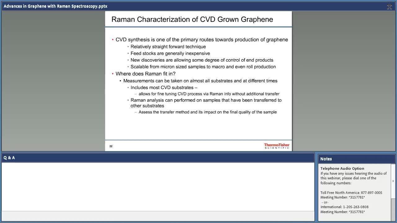
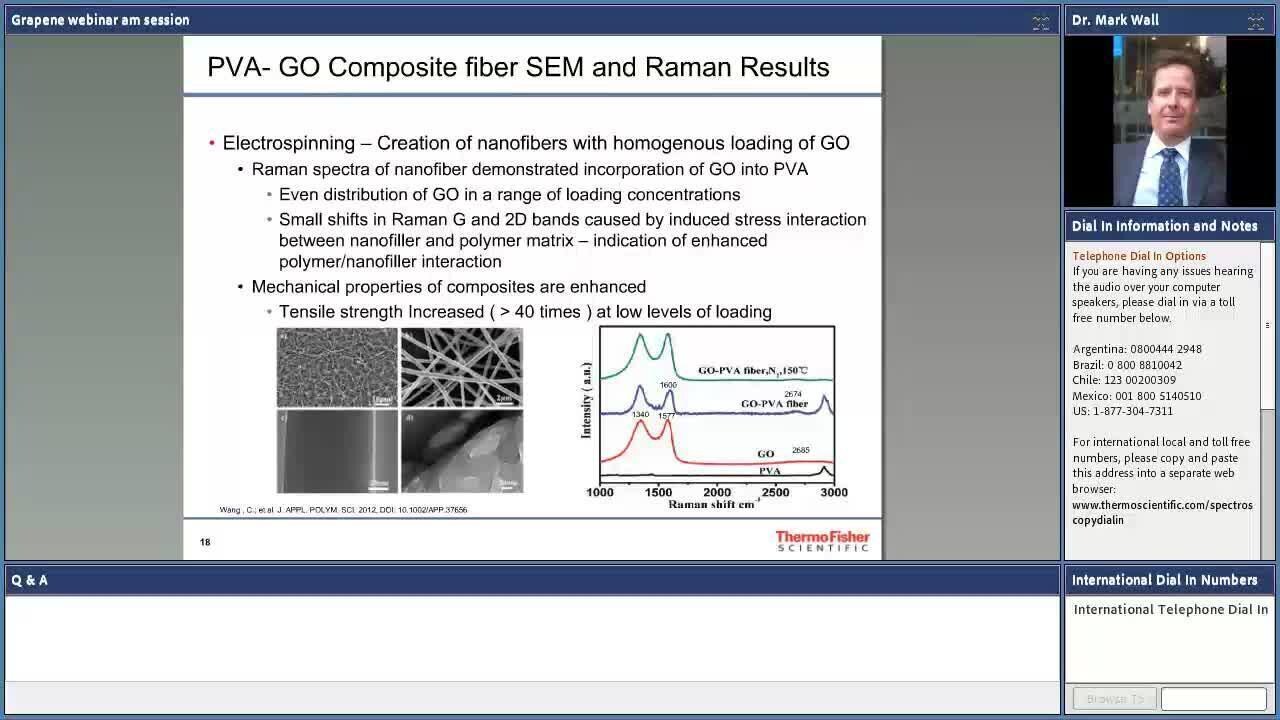
Graphene and based composites continue to be desirable materials due to mechanical, electrical and chemical properties. Potentially revolutionizing whole industries, from lighter- stronger advanced composites, flexible electronics, faster-smaller micro electronic devices, to more robust and efficient solar cell technologies, challenges associated with scaling up processes remain to be solved.
This webinar introduces how Raman imaging aids overcoming challenges associated with development of graphene and carbon nanotube-based technologies. We will cover:
- Utilize an image-centric approach to chemical imaging through an intuitive interface
- Simplify collection parameter set up and obtain instantaneous visual based results with real-time interpretation
- Specific examples presented will demonstrates how Raman imaging provides critical insight into graphene and based composite materials


Forensics
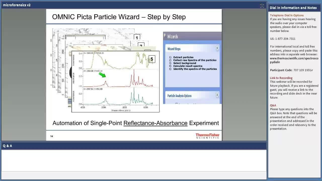
Trace evidence analysis: Raman and FTIR working together
Trace evidence examiners are required to obtain as much information as possible from microscopic amounts of material whilst respecting the need to maintain the integrity of the samples for any future forensic requirements. This webinar will demonstrate how spectroscopy plays a role in assisting the courts to answer some very difficult questions, with specific examples including additional levels of discrimination of fibers, glass; DNA –friendly methods of condom lubricant analysis and the potential for further discrimination of gun-shot residue.


Life sciences
Supplemental detection and classification methods of glaucoma disease states using Raman spectroscopy
This webinar discusses the potential of using Raman spectroscopy as a supplemental detection tool of glaucomatous changes in retinal tissues in vitro. Raman spectroscopic imaging was conducted on normal retinal tissues and retinal tissues with a variety of symptoms related to glaucoma. This webinar will address the use of Raman spectroscopy in clinical research, screening for the detection and characterization of early-stage glaucoma disease.
This webinar discusses:
- Collection of Raman spectral from normal retinal tissue, as well as tissue from retinas exhibiting glaucoma, elevated intraocular pressure, and compressive optic neuropathy
- Spectroscopic discrimination between each class of tissues, and the potential to harness these differences for detection and even prevention of disease


Minerals, geology, and gems
Raman and infrared microscopy of minerals and fluid inclusions
Infrared and Raman spectroscopy are convenient and information-rich analytical techniques for characterizing a wide variety of samples important in earth science. Combined with microscopy, vibrational spectra can identify specific minerals, characterize contents of fluid inclusions, and produce chemical images of complex mixtures. This presentation discusses how infrared and Raman complement other micro-techniques such as optical microscopy and SEM-EDS. We will show how Raman microscopy can deliver key data not possible by any other technique.

Raman spectroscopy can provide information that is complementary to other techniques, or it might be the only feasible method of analysis in a conventional laboratory. Learn how Raman spectroscopy can be used routinely to non-destructively study geological samples.
Topics covered will include:
- Rapid identification of unknown minerals with no need for special sample preparation
- Textural and mineralogical information about carbonate rocks
- Analysis of unexposed mineral and fluid inclusions

Pharmaceutical
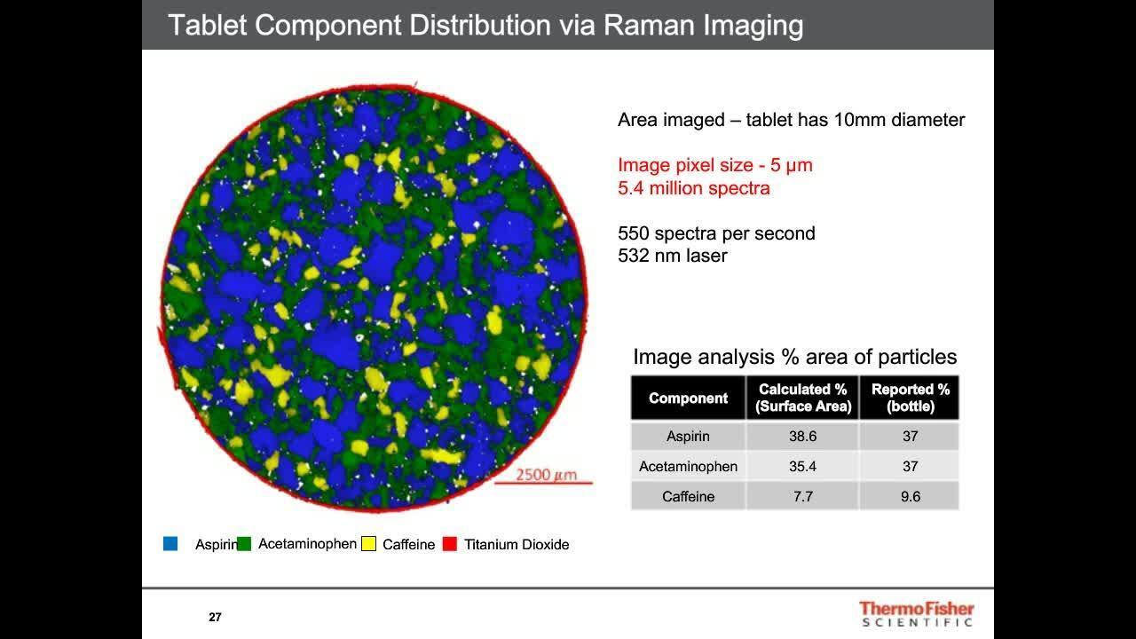
Visualizing, characterizing, and analyzing pharmaceutical constituents with Raman imaging
Pharmaceutical formations are typically complex mixtures that need to be carefully verified and understood. Raman spectroscopy is a proven method for identifying and verifying the presence of a variety of different components and providing detailed information on molecular structure and chemical environments. Raman imaging adds a spatial dimension to the analysis and extends the power of Raman spectroscopy across the sample.
Some of the applications of Raman imaging relevant to pharmaceutical evaluations include:
- Revealing spatial distribution of components - homogeneity and content uniformity
- Assessing particle size including relative percentages of components
- Differentiating and displaying chemically similar components such as polymorphs
- Quickly surveying larger areas and conducting detailed studies of smaller areas
Polymers
Your next packaging analysis tool – Raman microscopy
Raman microscopy is a valuable tool for packaging analysis, with special emphasis on food-contact materials. In minutes, you can characterize multilayer material properties such as structure and thickness and can even identify layers with minimal sample handling. In this webinar, we discuss novel promising applications of Raman microscopy, such as:
- Identification of migrants from adhesives through adjacent polymer or cardboard layers
- Detection of contaminants at ppm level in aqueous and oil food simulants after migration tests
- Simultaneous identification of several components of plastic materials such as base polymer, fillers, and stabilizers by means of software deconvolution tools.
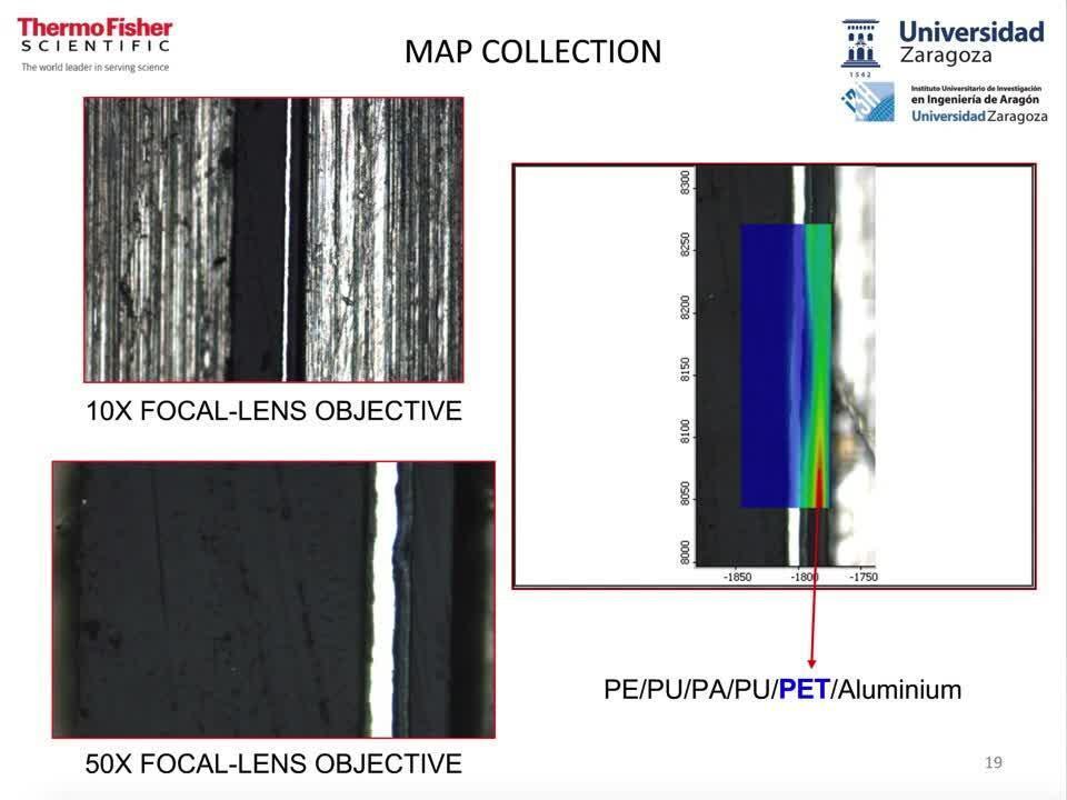
Raman microscopy is a powerful technique that can be utilized for the analysis of multilayer polymer films, thus enabling the control of composition and quality. Conventional Raman microscopy, which has spatial resolution as small as a micron, can be employed to analyze cross sections of multilayer polymer films. Confocal Raman microscopy can be used in situations where a reduction in sample preparation is desired, as it can generate depth profiles of the multilayer films, with no requirement for cross sectioning.
For Research Use Only. Not for use in diagnostic procedures.