Search Thermo Fisher Scientific

Invitrogen™
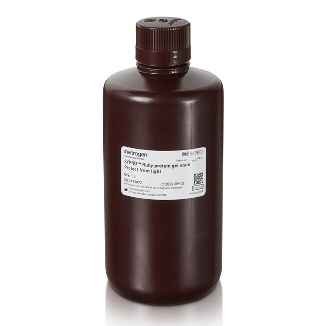
SYPRO™ Protein Gel Stains
SYPRO protein gel stains are sensitive, easy-to-use fluorescent stains for total proteins separated by polyacrylamide gel electrophoresis (PAGE).
| Catalog Number | Color | Quantity |
|---|---|---|
| S12000 | Ruby | 1 L |
| S6650 | Orange | 500 μL |
| S6651 | Orange | 10 x 50 μL |
| S12010 | Tangerine | 500 μL |
| S6653 | Red | 500 μL |
| S6654 also known as S-6654 | Red | 10 x 50 μL |
| S12001 | Ruby | 200 mL |
| S21900 | Ruby | 5 L |
| S12000X3 | Ruby | 3 x 1 L |
Catalog number S12000
Price (USD)
445.65
Online Exclusive
465.00Save 19.35 (4%)
Each
In stock
Color:
Ruby
Quantity:
1 L
Price (USD)
445.65
Online Exclusive
465.00Save 19.35 (4%)
Each
SYPRO protein gel stains are sensitive, easy-to-use fluorescent stains for total proteins separated by polyacrylamide gel electrophoresis (PAGE). Stained proteins can be viewed with a standard UV or blue-light transilluminator, or imaging equipment containing the appropriate filters or laser. SYPRO protein gel stains offer many advantages over Coomassie Blue and silver staining, including a fast, one-step staining protocol requiring no destaining, a linear dynamic range over three orders of magnitude, and very little protein-to-protein variation in staining.
SYPRO Ruby Protein Gel Stain features include:
• Convenient—ready-to-use formulation
• High sensitivity—detection to 0.25 to 1 ng
• Effective staining for 1D and 2D gels
• Can be multiplexed with other gel stains such as Pro-Q Diamond phosphoprotein gel stain or Pro-Q Emerald 300 glycoprotein gel stain
• Compatible with subsequent analysis of proteins by Edman-based sequencing or mass spectrometry
SYPRO Orange and SYPRO Red protein gel stain features include:
• Sensitive—detection to 4 to 8 ng
• Fast—rapid staining time of 10–60 min
• Best for 1D gels
• SYPRO Orange: Ex: 300, 470 nm, Em: 570 nm
• SYPRO Red: Ex: 300, 550 nm, Em: 630 nm
SYPRO Tangerine Protein Gel Stain features include:
• Sensitive—detection to 4 to 8 ng
• Fast—rapid staining time of 10 to 60 minutes
• Best for 1D gels
• Does not require fixation with acetic acid
• Compatible with subsequent gel staining, western blotting, zymography, electroelution or mass spectrometry
For Research Use Only. Not for use in diagnostic procedures.
Specifications
Concentration1X
Detection LocationIn-Gel Detection
Detection MethodFluorescence
Excitation/Emission280, 450/610 nm
Product LineSYPRO™
Product TypeProtein Gel Stain
Quantity1 L
Shelf Life9 Months
Shipping ConditionRoom Temperature
Target MoleculeProtein
ColorRuby
Label or DyeSYPRO Ruby
Unit SizeEach
Contents & Storage
SYPRO™ Ruby Protein Gel Stain is stable for at least 9 months when stored at room temperature and protected from light.

