Dynabeads™ Proteína G para inmunoprecipitación
Invitrogen™

Dynabeads™ Proteína G para inmunoprecipitación

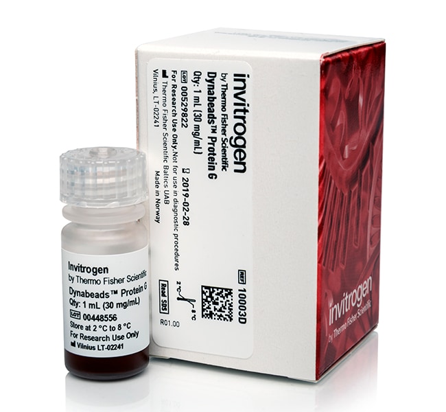
La proteína G Dynabeads son gránulos superparamagnéticos uniformes de 2,8 μm con proteína G recombinante (∼17 kDa) unida covalentemente aMás información
Have Questions?
Cambiar vistabuttonViewtableView
Número de catálogoCantidad
10003D1 ml
10004D5 ml
10009D50 mL
Número de catálogo 10003D
Precio (USD)
-
Cantidad:
1 ml
La proteína G Dynabeads son gránulos superparamagnéticos uniformes de 2,8 μm con proteína G recombinante (∼17 kDa) unida covalentemente a la superficie. La proteína G Dynabeads ofrece una alternativa superior a la Sepharose o la agarosa para la inmunoprecipitación (IP), y se dispone de protocolos manuales y automatizados.

• IP en menos de 40 minutos
• Alto rendimiento de proteínas objetivo con bajo consumo de anticuerpos
• Unión no específica muy baja con alta relación señal-ruido
• Sin necesidad de columnas, centrifugaciones ni larga limpieza previa
• Alta reproducibilidad y alto rendimiento compatible con instrumentos KingFisher

La separación manual de Dynabeads es rápida y fácil de realizar
El protocolo manual es sencillo y se puede realizar en menos de 40 minutos. En primer lugar, el anticuerpo de la proteína objetivo se incuba con la proteína G Dynabeads en un tubo durante 10 minutos. El exceso de anticuerpo se elimina colocando el tubo en un imán DynaMag y eliminando el sobrenadante. Los gránulos recubiertos de anticuerpos se pueden utilizar para una variedad de aplicaciones posteriores, como IP, Co-IP, IP de cromatina (ChIP), IP de ARN (RIP), purificación de IgG a pequeña escala y purificación de proteínas. El material unido se recopila fácilmente en un imán DynaMag gracias a las exclusivas propiedades magnéticas de Dynabeads. La proteína G recombinante de los gránulos no contiene puntos de unión de albúmina, por lo que la albúmina no se copurifica durante el procedimiento. El IP es rápido y ofrece un alto rendimiento, una alta reproducibilidad y una unión no específica muy baja, por lo que no es necesario realizar una limpieza previa.

La separación automatizada de Dynabeads ayuda a aumentar el rendimiento y reduce el tiempo de manipulación
Si está trabajando con varias muestras en paralelo, el número de pasos de lavado y el tiempo de manipulación aumenta proporcionalmente con el número de muestras. El pipeteo y otras manipulaciones manuales tienden a ser menos uniformes que la automatización cuando se trabaja con muchas muestras a la vez. Para manejar mejor un número de muestras de medio a alto rendimiento, reducir el tiempo de manipulación y asegurar una alta reproducibilidad, hemos desarrollado protocolos IP para los instrumentos KingFisher Flex y KingFisher Duo Prime. Los protocolos automatizados replican los protocolos manuales, que proporcionan un rendimiento proteico objetivo igual de alto y la misma unión no específica baja y alta reproducibilidad. No importa si está trabajando con 10 o 96 muestras, el protocolo IP es inferior a 40 minutos, independientemente. Solo tiene que cargar los reactivos en las placas, pulsar el botón de “inicio” y, para cuando haya preparado el análisis posterior, la IP estará lista. Es posible que sea necesaria cierta optimización (por ejemplo, los tiempos de incubación) dependiendo de su anticuerpo y de la abundancia y/o especificidad de su proteína objetivo.

• Utilice el instrumento KingFisher Duo para un rendimiento bajo a medio (1-12 muestras/ciclo)
• Utilice el instrumento KingFisher Flex para un rendimiento alto (12-96 muestras/ciclo)
Ver protocolos automatizados
Ver un vídeo sobre el instrumento KingFisher Flex

La separación suave provoca un estrés físico mínimo en las proteínas
La tecnología de separación magnética que utiliza la proteína G Dynabeads es rápida y suave, de forma que causa el mínimo estrés físico en las proteínas objetivo. Esto permite el aislamiento y la concentración de compuestos lábiles que, de otro modo, podrían disociarse o quedar dañados por las proteasas durante largos tiempos de incubación. Se preserva la conformación de proteínas nativas y los grandes complejos de proteínas.

Fuerza y capacidad de unión
La proteína G Dynabeads permite el aislamiento de la mayoría de las inmunoglobulinas de mamíferos (Ig). La cantidad de Ig capturada depende de la concentración de Ig en la muestra inicial, así como del tipo y la fuente de Ig. 100 μl de proteína G Dynabeads aislarán aproximadamente 25–30 μg de IgG humana a partir de una muestra que contiene 20–200 μg/ml de IgG. La unión Fc predominante permite una orientación de Ig óptima. Los anticuerpos se unen a la superficie lisa exterior de los gránulos, de modo que no quedan atrapados en poros grandes, como sucede con los gránulos basados en Sepharose/agarosa. Todos los anticuerpos están disponibles para la unión de proteínas, por lo que se requieren cantidades bajas de anticuerpos mientras se sigue obteniendo el mismo alto rendimiento de la proteína diana. La superficie lisa del gránulo también es responsable de la unión baja no específica por la que son conocidos los Dynabeads.

Obtenga más información acerca de Dynabeads
•La Proteína A Dynabeads también está disponible como un kit listo para usar con tampones incluidos
Ver guías de selección de inmunoprecipitación, datos y referencias
Ver imanes para separaciones de Dynabeads
Buscar productos Dynabeads para otras aplicaciones

Comprar en OEM
Para comprar Proteína A y Proteína G Dynabeads en un OEM, póngase en contacto con nuestro departamento de ventas de OEM y de licencias de productos.

*Sepharose es una marca comercial de GE Healthcare Bio-Sciences AB.
Para uso exclusivo en investigación. No apto para uso en procedimientos diagnósticos.

Especificaciones
Certificaciones/conformidadISO9001 and ISO13485
Concentración30 mg/mL
DescripciónRecombinant Protein G covalently bound to Dynabeads
Diámetro (métrico)2,8 μm
Para utilizar con (aplicación)Inmunoprecipitación
Para utilizar con (equipo)KingFisher™ Duo Prime, KingFisher™ Flex
FormatoMicroesferas en suspensión
Compatibilidad de alto rendimientoCompatible con alto rendimiento
Tipo de ligandoProteína G
MaterialPoliestireno
Grado de pureza o calidadFor Research Use Only
Cantidad1 ml
Duración de almacenamiento24 meses a partir de la fecha de fabricación
Condiciones de envíoTemperatura ambiente
Suficiente para20 Tests
Funcionalidad de superficieProtein G
ObjetivoAntibodies
UniformidadMonosized 2.8 mm (CV <5%)
Línea de productosDynabeads™
TipoGránulo superparamagnético de proteína G
Unit SizeEach
Contenido y almacenamiento
Contiene: 1 ml de gránulos Dynabeads™ con proteína G
Almacenamiento: 2 °C a 8 °C.

Preguntas frecuentes

My Dynabeads magnetic beads are not pelleting well with the magnet. Do you have any suggestions for me?

Please review the following possibilities for why your Dynabeads magnetic beads are not pelleting:

- The solution is too viscous.
- The beads have formed aggregates because of protein-protein interaction.

Try these suggestions: - Increase separation time (leave tub on magnet for 2-5 minutes)
- Add DNase I to the lysate (~0.01 mg/mL)
- Increase the Tween 20 concentration to ~0.05% of the binding and/or washing buffer.
- Add up to 20 mM beta-merecaptoethanol to the binding and/or wash buffers.

Find additional tips, troubleshooting help, and resources within our Dynabeads Nucleic Acid Purification Support Center.

I have a long double-stranded DNA fragment I would like to isolate. What product do you recommend?

For biotin-labeled DNA that is less than 1 kb, we recommend you use Dynabeads M270 Streptavidin (Cat. No. 65305) and MyOne C1 magnetic beads (Cat. No. 65001). We recommend our Dynabeads KilobaseBINDER Kit (Cat. No. 60101), which is designed to immobilize long (>1 kb) double-stranded DNA molecules. The KilobaseBINDER reagent consists of M-280 Streptavidin-coupled Dynabeads magnetic beads along with a patented immobilization activator in the binding solution to bind to long, biotinylated DNA molecules for isolation. Please see the following link (https://www.thermofisher.com/us/en/home/life-science/dna-rna-purification-analysis/napamisc/capture-of-biotinylated-targets/immobilisation-of-long-biotinylated-dna-fragments.html) for more information in regards to long biotinylated DNA fragment isolation.

Find additional tips, troubleshooting help, and resources within our Dynabeads Nucleic Acid Purification Support Center.

Can I use Dynabeads magnetic beads to isolate single-stranded DNA templates?

Yes, Dynabeads magnetic beads can be used to isolate single-stranded DNA. Streptavidin Dynabeads magnetic beads can be used to target biotinylated DNA fragments, followed by denaturation of the double-stranded DNA and removal of the non-biotinylated strand. The streptavidin-coupled Dynabeads magnetic beads will not inhibit any enzymatic activity. This enables further handling and manipulation of the bead-bound DNA directly on the solid phase. Please see the following link (https://www.thermofisher.com/us/en/home/life-science/dna-rna-purification-analysis/napamisc/capture-of-biotinylated-targets/preparing-single-stranded-dna-templates.html) for more information in regards to single-stranded DNA capture.

Find additional tips, troubleshooting help, and resources within our Dynabeads Nucleic Acid Purification Support Center.

What is the magnetic susceptibility for Dynabeads magnetic beads?

Magnetic susceptibility is a measure of how quickly the beads will migrate to the magnet. This will depend on the iron content and the character of the iron oxide. The magnetic susceptibility given for the Dynabeads magnetic beads is the mass susceptibility, given either as cgs units/g or m^3/kg (the latter being an SI unit). For ferri- and ferromagnetic substances, the magnetic mass susceptibility is dependent upon the magnetic field strength (H), as the magnetization of such substances is not a linear function of H but approaches a saturation value with increasing field. For that reason, the magnetic mass susceptibility of the Dynabeads magnetic beads is determined by a standardized procedure under fixed conditions. The magnetic mass susceptibility given in our catalog is thus the SI unit. Conversion from Gaussian (cgs, emu) units into SI units for magnetic mass susceptibility is achieved by multiplying the Gaussian factor (emu/g or cgs/g) by 4 pi x 10^-3. The resulting unit is also called the rationalized magnetic mass susceptibility, which should be distinguished from the (SI) dimensionless magnetic susceptibility unit. In general, magnetic mass susceptibility is a measure of the force (Fz) influencing an object positioned in a nonhomogenous magnetic field. The magnetic mass susceptibility of the Dynabeads magnetic beads is measured by weighing a sample, and then subjecting the sample to a magnetic field of known strength. The weight (F1) is then measured, and compared to the weight of the sample when the magnetic field is turned off (F0). The susceptibility is then calculated as K x 10^-3 = [(F1-F0) x m x 0.335 x 10^6], where K is the mass susceptibility of the sample of mass m. The susceptibility is then converted to SI units.

Find additional tips, troubleshooting help, and resources within our Dynabeads Nucleic Acid Purification Support Center.

How can I determine coupling efficiency of Dynabeads magnetic beads?

There are different methods to check binding of ligands to the beads, including optical density (OD) measurement, fluorescent labeling, and radioactive labeling.

For OD measurement, you would measure the OD of the ligand before immobilization to the beads and compare it with the ligand concentration that is left in the supernatant after coating. This gives a crude measurement of how much protein has bound to the beads.

Protocol:

1.Set spectrophotometer to the right wavelength. As a blank, use the Coupling Buffer.
2.Measure the absorbance of the Pre-Coupling Solution. A further dilution may be necessary to read the absorbance, depending upon the amount of ligand added.
3.Measure the absorbance of the Post-Coupling Solution. A dilution may be necessary to read the absorbance.
4.Calculate the coupling efficiency, expressed as the % protein uptake, as follows. [(Pre-Coupling Solution x D) - (Post-Coupling Solution x D)] x 100/(Pre-Coupling Solution x D) where D = dilution factor.

For fluorescent labeling, we suggest negatively quantifying the amount of ligand bound by measuring ligand remaining in the coupling supernatant (compared to the original sample), rather than directly measuring the ligands on the beads. Add labeled ligand to the beads, and measure how much ligand is left in the supernatant (not bound to the beads). By comparing this with the total amount added in the first place, you can then calculate how much of the ligand that has been bound to the beads. Keep in mind that the Dynabeads magnetic beads are also autofluorescent, which is why direct measuring of fluorescence of the bead-bound ligands is not recommended, but rather this indirect approach. The label could be, for example, FITC/PE. Some researchers perform a direct approach with success (using a flow cytometer).

Radioactive labeling is the most sensitive method of the three, but it is also the most difficult one. It involves radioactively labeling a portion of the ligand. We use radiolabeled I-125 in tracer amounts and mix it with "cold" ligands in a known ratio before coupling. The absolute quantities for the ligand on the beads should be obtained by measuring the beads in a scintillation (gamma) counter and comparing the cpm with a standard.

Protocol:

1.Take out an appropriate amount of beads and wash the beads in 1 mL of binding buffer.
2.Pipette out desired amount of human IgG in a separate tube.
3.Mix the human IgG with I-125-labeled human IgG (30,000 - 100,000 cpm).
4.Dilute the mixture of human IgG and I-125-labeled human IgG to 100 mL in binding buffer.
5.Incubate for 30 minutes at room temperature and measure the cpm in a scintillation counter.
6.Wash the beads (with coating) four times, and measure cpm again.
The % binding is calculated by using the equation : (cpm after washing/cpm before washing)x100%.

Find additional tips, troubleshooting help, and resources within our Dynabeads Nucleic Acid Purification Support Center.

Documentos y descargas

Certificados

Hojas de datos de seguridad

Compartir número de catálogo, nombre o enlace.