Search Thermo Fisher Scientific
제품 개요
그림
동영상
관련 제품 추천
관련 제품 추천
문서
FAQ
인용 및 참조 문헌
추가 정보
관련 제품 추천
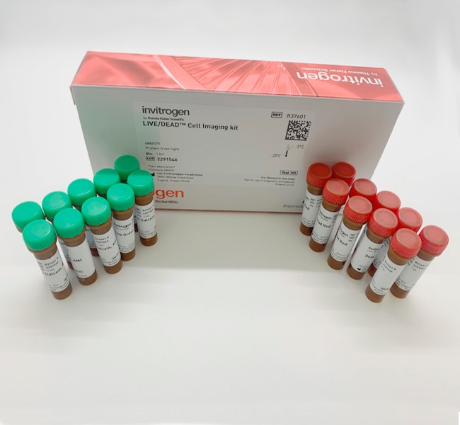
The LIVE/DEAD® Cell Imaging Kit is a sensitive two-color fluorescence cell viability assay optimized for FITC and Texas Red™ filters. Quick and easy to use, the kit allows discrimination between live and dead cells with two probes that measure recognized parameters of cytotoxicity and cell viability—intracellular esterase activity and plasma membrane integrity.
Contains Calcein, AM, cell permeant dye as live cell indicator and BOBO-3 Iodide as dead cell indicator.
See other Ready Probes ready-to-use imaging reagents and accessories ›
The LIVE/DEAD® Cell Imaging Kit *488/570* offers:
Fast, simple determination of live and dead cells
• Accuracy with convenience
• Sensitive probes ideal for FITC and Texas Red filters
Dual Probe-Based Assay for Imaging Platforms
Just like our popular LIVE/DEAD® Viability/Cytotoxicity assay, the new LIVE/DEAD® Cell Imaging Kit is based on a cell-permeable dye for staining of live cells and a cell-impermeable dye for staining of dead and dying cells, which are characterized by compromised cell membranes. To adapt this important assay for imaging platforms, the LIVE/DEAD® Cell Imaging Kit components were optimized for the common green and red imaging filters used with FITC and Texas Red. The live cell component produces an intense, uniform green fluorescence in live cells (ex/em 488 nm/515 nm). The dead cell component produces a predominantly nuclear red fluorescence (ex/em 570nm/602 nm) in cells with compromised cell membranes, a strong indicator of cell death and cytotoxicity (Fig 1).
Quick, Exact Determination of Viability and Cytotoxicity
The LIVE/DEAD® Cell Imaging kit provides very sensitive detection of key measures of cell health: viability, live/dead ratio, and cytotoxicity. The components of the LIVE/DEAD® Cell Imaging kit are configured for ease of use with minimal dilutions.
Assay Principle
With the LIVE/DEAD® Cell Imaging Kit, live cells are distinguished by the presence of ubiquitous intracellular esterase activity as determined by the enzymatic conversion of the virtually non-fluorescent cell-permeant calcein AM to the intensely fluorescent calcein, which is well-retained within live cells. The red component of the LIVE/DEAD® Cell Imaging Kit is cell-impermeant and therefore only enters cells with damaged membranes. In dying and dead cells a bright red fluorescence is generated upon binding to DNA. Background fluorescence levels are inherently low with this assay technique because the dyes are virtually non-fluorescent before interacting with cells. The fluorophores in the LIVE/DEAD® Cell Imaging Kit were selected for their brightness, spectral properties (FITC and Texas Red filters), and ease of use. Packaged for workflow convenience, they allow for effortless and quick determination of the fractions of live and dead cells in a population.
See other Molecular Probes imaging tools and reagents
사양
세포 유형
Mammalian Cells
색상
Red, Green
설명
LIVE/DEAD™ Cell Imaging Kit (488/570)
검출 방법
Fluorescence
용도(애플리케이션)
Viability Assay
용도 (장비)
Floid Cell Imaging System, Fluorescence Microscope
제품 유형
Cell Imaging Kit
염료 유형
Other Label(s) or Dye(s)
방출
515, 602 nm
여기 파장 범위
488, 570 nm
형태
Frozen
형식
Tube(s)
인큐베이션 시간
15 min
제품라인
LIVE/DEAD™, Molecular Probes™
수량
1 mL
배송 조건
Dry Ice
구성 및 보관
• Component A: 10 x 1 mL vials
• Component B: 1 x 10 vials (dried down)
Store at -20°C
• Component B: 1 x 10 vials (dried down)
Store at -20°C
그림
문서 및 다운로드
Certificates | 증명서
Lot 번호 또는 부분 Lot 번호로 검색하십시오.
자주 묻는 질문(FAQ)
인용 및 참조 문헌
Search citations by name, author, journal title or abstract text
