Powerful, multifaceted 2D–5D software for visualization, processing and analysis of microscopy imaging serving drug discovery
Supporting scientists and imaging experts on the challenging path toward drug discovery need state-of-the-art, repeatable, and automated 2D/3D software solutions. Thermo Scientific Amira Software reduces manual steps in the drug discovery process while increasing accuracy, time, and cost-effectiveness. From target discovery to compound identification, Amira Software is a powerful, multi-faceted 2D-5D platform for visualizing, manipulating, and understanding life science research data from many image modalities, including CT (Computed Tomography), MRI (Magnetic Resonance Imaging), 3D microscopy, and other techniques. It offers a comprehensive toolbox of image processing methods that can be automated through custom detection workflows.
Life science researchers continue to drive drug discovery and academic research down to a structural, sub-cellular level using automated, all-in-one tools and technologies. In pushing these limits, scientists can better understand how cells function and respond to disease or genetic variations, using their findings to advance research and development for breakthrough drug discoveries.
Imaging data
Processing
Visualization
Analysis
Empower your lab with powerful and comprehensive imaging data analysis toolbox
Amira Software offers high-end processing, registration, segmentation, visualization, animation, and data analysis to support your advanced discovery workflows in pharmaceutical research areas ranging from structural and cellular biology to tissue imaging, neuroscience, preclinical imaging, and bioengineering.
Accelerate time to data and time to market
By providing ready-to-use recipes and AI-powered automated processing tools, Amira Software supports faster image analysis reducing the number of manual steps. The software has built-in support for deep learning models and includes a ready-to-use Python environment. By creating automated workflows, Amira Software supports scientists with faster image analysis and reproducibility, reducing the learning curve for inexperienced users. Deep learning models accelerate and improve the segmentation and interpretation of complex or large data sets.
Analyze and correlate data from any imaging modality, at any scale
Amira Software offers native compatibility and expansive correlative analysis to help you better understand your 2D-5D imaging data. It is a flexible software solution that can handle any modality at any scale and size. With a multi-volume infrastructure, Amira Software is designed to perform image registration on as many modalities as desired. It can also perform correlative analysis at either single points in time or as part of longitudinal studies.
Confidence
Thanks to powerful segmentation and image processing capabilities and workflows, and after 20+ years of collaboration with the scientific community and thousands of researchers, it has been now proven that our digital imaging-based workflows provide reliable answers to biomedical and life science research.
Support
Thanks to our dedicated professional support team, you get access to our top experts to ensure that no question is left unanswered. And with our training, consulting options, and ever-expanding collection of tutorials and how-to’s, you can reduce your learning curve and focus on getting the answers you are pursuing.
Customization
Because your needs are unique and keep evolving, our software solutions are fully flexible and open to customization. Thanks to our scripting interface (Python, TCL), bridge with MATLAB, and our programming API, you can expand our software solution and integrate your own IP (Intellectual Property). And, if needed, our professional service team can help you design unique solutions tailored to your needs.
With its long-standing applications in multi-modality imaging techniques, Amira Software is the tool of choice for many researchers working with data from different imaging modalities. Amira architecture supports multi-volume data processing and visualization from the ground up. The automatic registration tools integrated into the standard edition of Amira Software with the Multi-Planar Workroom offer the flexibility of the most commonly used automated registration metrics and optimizers that allow for the adjustment of the registration parameters to meet the needs of your data.
Membrane detection
Amira Software accelerates segmentation by employing automated detection of cellular features, such as filaments and membranes. By performing advanced image analysis and statistics on the cryo-tomograms, Amira Software combines data from these individual structures, averaging out noise and increasing contrast to create a composite model with higher resolution.
Cell detection
Cellular spheroids are 3D cell cultures that represent in vivo tissue better than traditional cellular monolayers, improving the ability of researchers to study cancer, diabetes mellitus, or stem cells among many applications. However, cell analysis in such 3D multicellular objects is not accurately possible, especially without destroying the object of study. Amira Software can automatically separate the cells and perform volumetric, morphometric, and intensity measurements on individual cells and their nuclei in 3D image data of these spheroids. Amira Software also helps researchers with the analysis of conventional 2D cellular monolayers.
High Content Screening
Amira Software enables multi-channel quantitivative capabilities to investigate the most demanding scientific and experimental models used in HCS, including immuno-oncology, neuroscience, and organoid health. Thanks to its incredible speed and flexibility, Amira Software helps enable advanced 2D–5D bioimaging workflows in research areas ranging from structural and cellular biology, to neuronal and spheroid morphology in 3D models. To facilitate these advanced analyses, Amira Software features deep learning for improved training and prediction, including "intelligent denoising functionality
With Amira Software’s new Xplore5D extension, life scientists can easily visualize, correlate, and process large multi-channel and time series data in one platform, without worrying about image size or memory limits. With its smart file format, Xplore5D allows you to compress all imaging data without losing precious information collected during acquisition.
With Amira Software, you are able to use a template-matching algorithm to automatically detect and trace these fine filaments in noisy cryo-EM or DualBeam data. In addition, Amira Software enables you to reconstruct filamentous networks and edit the resulting graphs to remove image features erroneously identified as filaments or to add missing parts of a network. Graphs of filaments can then be quantified by measuring parameters such as amount, length, thickness, orientation, ranks, etc. Compelling 3D renderings can be created from the reconstructed model and the computed parameters.
With its long-standing applications in multi-modality imaging techniques, Amira Software is the tool of choice for many researchers working with data from different imaging modalities. Amira architecture supports multi-volume data processing and visualization from the ground up. The automatic registration tools integrated into the standard edition of Amira Software with the Multi-Planar Workroom offer the flexibility of the most commonly used automated registration metrics and optimizers that allow for the adjustment of the registration parameters to meet the needs of your data.
Amira Software accelerates segmentation by employing automated detection of cellular features, such as filaments and membranes. By performing advanced image analysis and statistics on the cryo-tomograms, Amira Software combines data from these individual structures, averaging out noise and increasing contrast to create a composite model with higher resolution.
Cellular spheroids are 3D cell cultures that represent in vivo tissue better than traditional cellular monolayers, improving the ability of researchers to study cancer, diabetes mellitus, or stem cells among many applications. However, cell analysis in such 3D multicellular objects is not accurately possible, especially without destroying the object of study. Amira Software can automatically separate the cells and perform volumetric, morphometric, and intensity measurements on individual cells and their nuclei in 3D image data of these spheroids. Amira Software also helps researchers with the analysis of conventional 2D cellular monolayers.
Amira Software enables multi-channel quantitivative capabilities to investigate the most demanding scientific and experimental models used in HCS, including immuno-oncology, neuroscience, and organoid health. Thanks to its incredible speed and flexibility, Amira Software helps enable advanced 2D–5D bioimaging workflows in research areas ranging from structural and cellular biology, to neuronal and spheroid morphology in 3D models. To facilitate these advanced analyses, Amira Software features deep learning for improved training and prediction, including "intelligent denoising functionality
With Amira Software’s new Xplore5D extension, life scientists can easily visualize, correlate, and process large multi-channel and time series data in one platform, without worrying about image size or memory limits. With its smart file format, Xplore5D allows you to compress all imaging data without losing precious information collected during acquisition.
With Amira Software, you are able to use a template-matching algorithm to automatically detect and trace these fine filaments in noisy cryo-EM or DualBeam data. In addition, Amira Software enables you to reconstruct filamentous networks and edit the resulting graphs to remove image features erroneously identified as filaments or to add missing parts of a network. Graphs of filaments can then be quantified by measuring parameters such as amount, length, thickness, orientation, ranks, etc. Compelling 3D renderings can be created from the reconstructed model and the computed parameters.
Services
Introductory training
Shorten your learning curve and maximize your investment with this introductory training specifically designed for new users of Amira, Avizo and PerGeos Software.
The course consists of a lecture with hands-on sessions. The training material highlights the basic features and functionalities of Amira, Avizo and PerGeos Software.
Advanced training
Maximize your investment and reduce your time-to-results with this advanced training specifically designed for existing users of Amira, Avizo and PerGeos Software.
The course consists of a lecture with hands-on sessions. The training material highlights advanced features and functionalities of Amira, Avizo and PerGeos Software.
Custom development
With over 25 years of experience in 3D and image processing and hundreds of custom projects delivered to organizations small and large, Thermo Fisher Scientific can provide you with a solution tailored to fit your specific needs.
We can customize and expand our software solutions at various levels.
Beginning of dialog window. Escape will cancel and close the window.
End of dialog window.
This is a modal window. This modal can be closed by pressing the Escape key or activating the close button.
This is a modal window. This modal can be closed by pressing the Escape key or activating the close button.
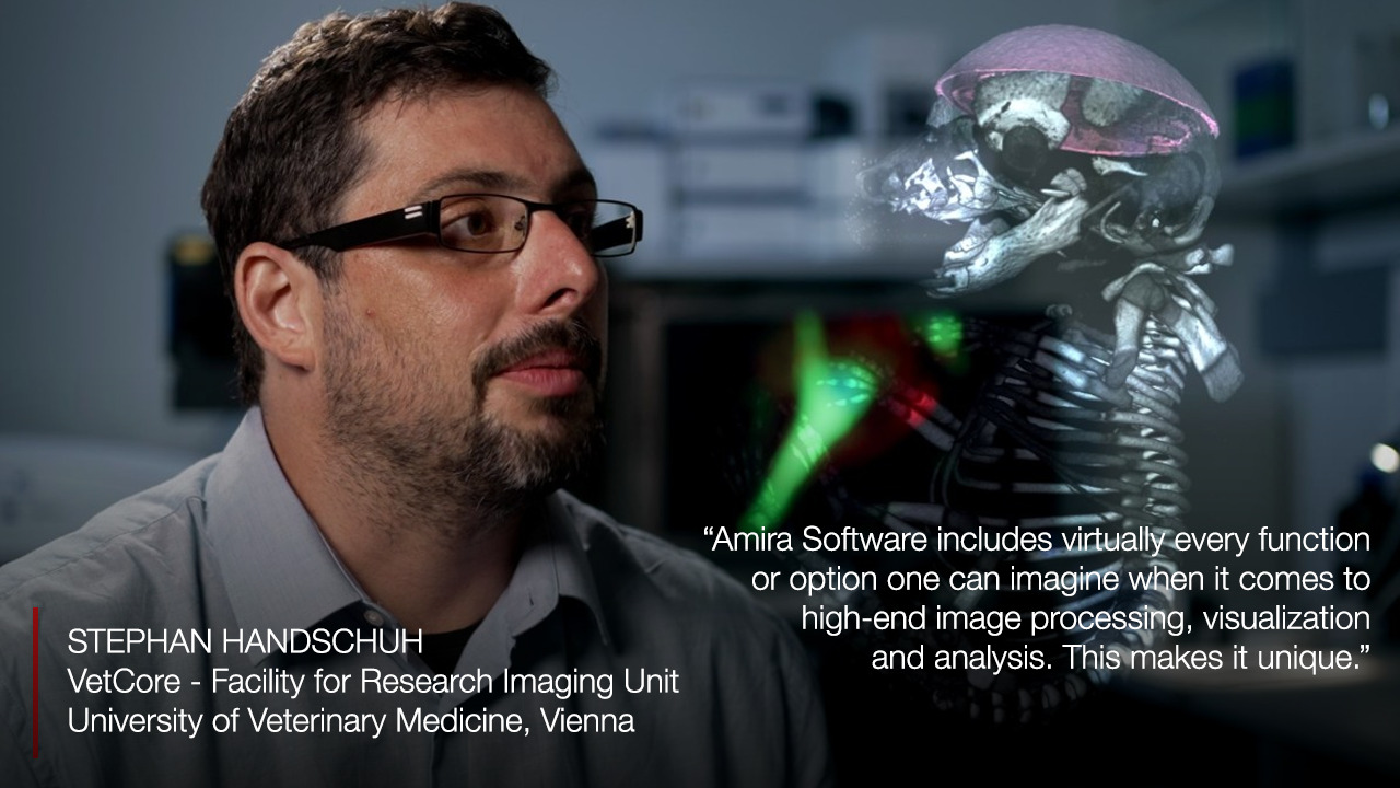
VetCore, University of Veterinary Medicine uses Amira Software
"Research at Vetmeduni Vienna is quite diverse, covering both clinical and preclinical research areas. We are currently four people working in the imaging facility, providing equipment and supports for researchers in both image acquisition (widefield microscopy, CLSM, spinning disk confocal microscopy, soon also STORM super-resolution microscopy, microscopic X-ray computed tomography) and in data visualization and analysis. Amira Software allows you to build networks, enabling complex interactions between data objects. Biology is also complex, and biological interactions are complex networks. Complex questions require complex tools to address them. In this respect, analysis networks such as used in Amira Software outperform most other software approaches when it comes to complex data analysis. Furthermore, I like that Amira Software is quite open concerning integration of deep learning networks and scripting (Tcl, Matlab, Python,...)."
Stephan Handschuch, PhD VetCore, University of Veterinary Medicine, Vienna
Video Player is loading.
Current Time 0:00
/
Duration 6:00
Loaded: 2.73%
0:00
Stream Type LIVE
Remaining Time -6:00
1x
Chapters
descriptions off, selected
captions settings, opens captions settings dialog
captions off, selected
en (Main), selected
This is a modal window.
Beginning of dialog window. Escape will cancel and close the window.
End of dialog window.
This is a modal window. This modal can be closed by pressing the Escape key or activating the close button.
This is a modal window. This modal can be closed by pressing the Escape key or activating the close button.
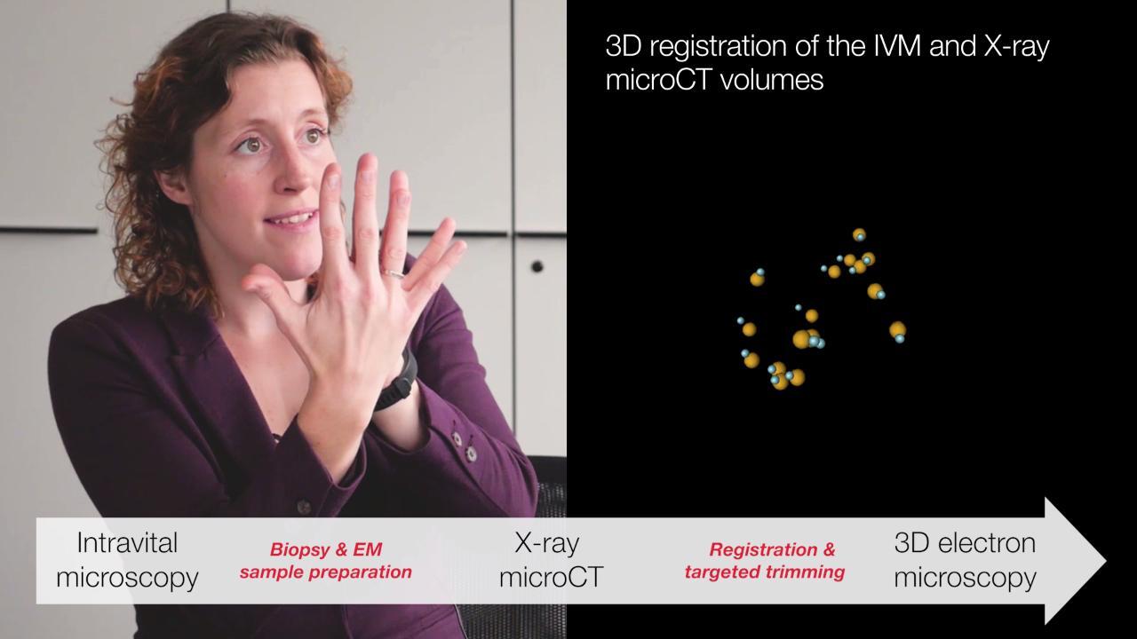
EMBL and DKFZ uses Amira Software
The present testimonial highlights the way Amira Software supported Matthia A. Karreman, MSc, PhD at EMBL and DKFZ, Heidelberg, Germany, in her project on multimodal correlative microscopy to efficiently target single tumor cells in vivo. Read reference paper. Video: 6 min
- Bio-Formats - Bitmap formats - Microscopy: electron and optical - Medical and neuroimage formats - Molecular formats - Other acquisition devices (MRI, radiography, etc.)
Finite element modeling, geometric modeling, CAD
Support for multi-data/multi-view, multi-channel, time series, very large data
Scaling, calibration, conversion, re-sampling
Image enhancement, comprehensive filtering and convolution, Fourier frequency transforms