Build your flow cytometry panels for FREE with our scientists using our Panel Design Service or utilize our Panel Builder Tool to self-build your panels. We provide options for all experience levels when it comes to designing your flow cytometry panel.
Getting started with our free panel design service
We have a team of technical support scientists available to help you. Our team provides this Panel Design Service for those who want help with building their panel every step of the way.
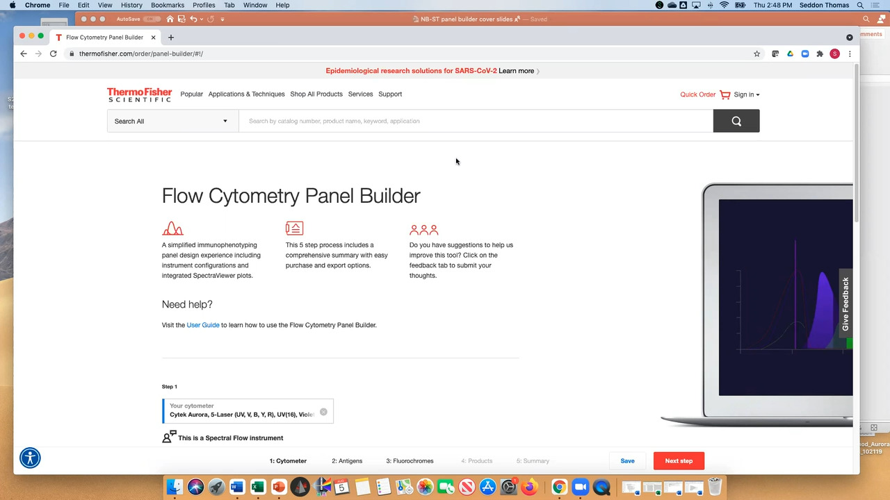
Getting started with the flow cytometry panel builder tool
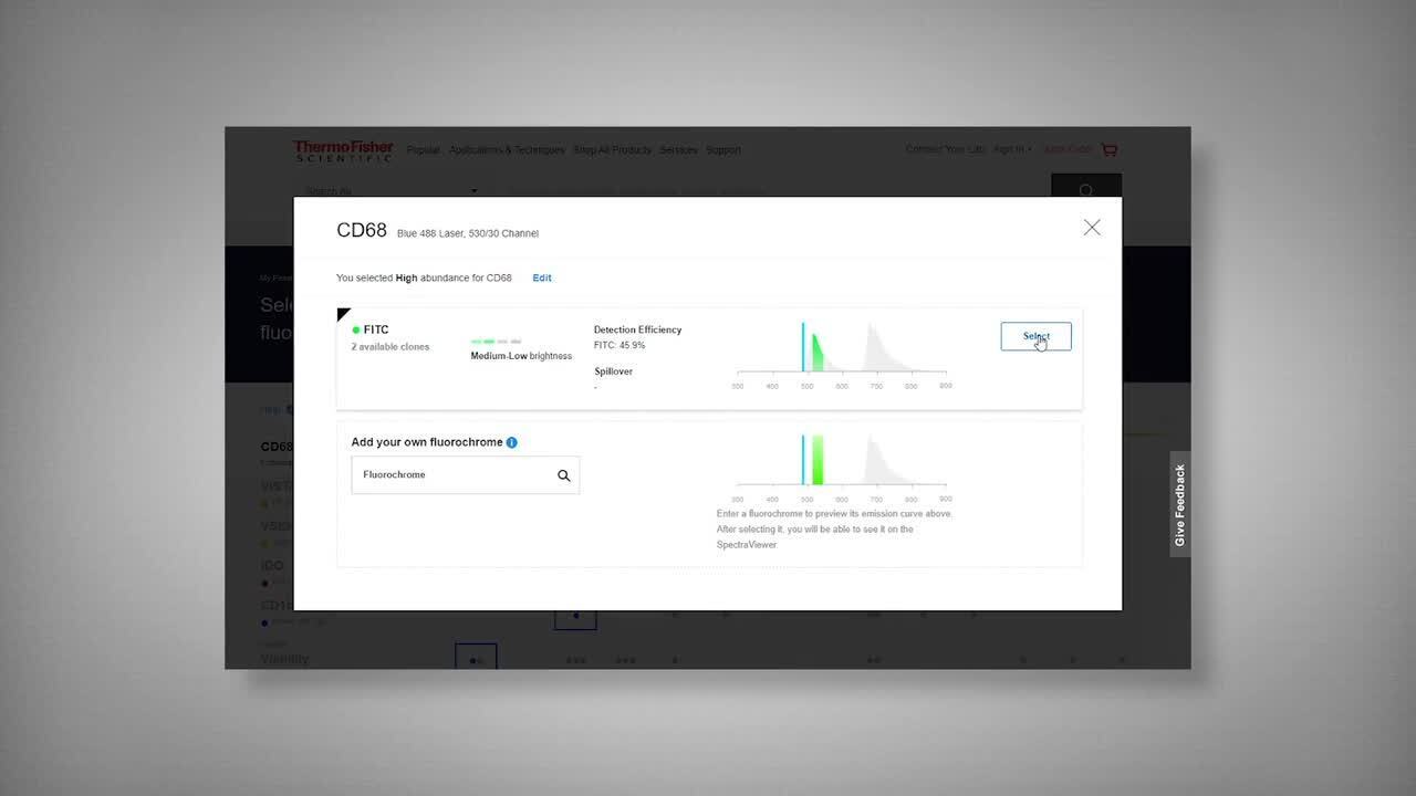
Choosing the optimal combination of fluorochromes can be simplified with a guided method. The Flow Panel Builder offers a curated and customizable approach to the panel building process.
Get your flow cytometry panels designed in 4 easy steps
Click image to enlarge
Be the spark story - Berencia
Inspiring flow cytometry scientists, we create new multicolor panels at no cost for you.
Video Player is loading.
Current Time 0:00
/
Duration 3:16
Loaded: 0%
0:00
Stream Type LIVE
Remaining Time -3:16
1x
Chapters
descriptions off, selected
captions settings, opens captions settings dialog
captions off, selected
en (Main), selected
This is a modal window.
Beginning of dialog window. Escape will cancel and close the window.
End of dialog window.
This is a modal window. This modal can be closed by pressing the Escape key or activating the close button.
This is a modal window. This modal can be closed by pressing the Escape key or activating the close button.
Getting started with the flow cytometry panel builder tool
Choosing the optimal combination of fluorochromes can be simplified with a guided method. The Invitrogen Flow Panel Builder offers a customizable panel building process to fit your flow cytometry experimental needs, whatever your experience level.
Video Player is loading.
Current Time 0:00
/
Duration 8:21
Loaded: 0.00%
0:00
Stream Type LIVE
Remaining Time -8:21
1x
Chapters
descriptions off, selected
captions settings, opens captions settings dialog
captions off, selected
en (Main), selected
This is a modal window.
Beginning of dialog window. Escape will cancel and close the window.
End of dialog window.
This is a modal window. This modal can be closed by pressing the Escape key or activating the close button.
This is a modal window. This modal can be closed by pressing the Escape key or activating the close button.
Video: how to use the panel builder
Watch the video to learn how to use the Invitrogen Flow Cytometry Panel Builder to build your next flow cytometry panel in 5 easy steps.
Video Player is loading.
Current Time 0:00
/
Duration 14:02
Loaded: 0%
00:00
Stream Type LIVE
Remaining Time -14:02
1x
Chapters
descriptions off, selected
captions settings, opens captions settings dialog
captions off, selected
en (Main), selected
This is a modal window.
Beginning of dialog window. Escape will cancel and close the window.
End of dialog window.
This is a modal window. This modal can be closed by pressing the Escape key or activating the close button.
This is a modal window. This modal can be closed by pressing the Escape key or activating the close button.
Video: how to design spectral flow cytometry experiments
Watch the video to create a panel for your spectral instrument
List of available fluorophores based on their usage, benefits, and intended applications with Flurophore Selection Guide.
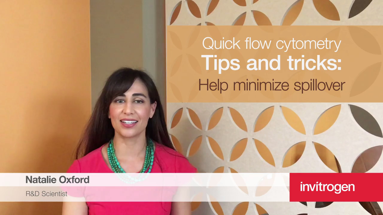
Any time you have markers that you know will be co-expressed on your cells of interest, make sure to space them out into separate channels. If you will need to use any adjacent channels, that's where you would put any markers that are mutually exclusive so that they'll still be easy to distinguish.
Video Player is loading.
Current Time 0:00
/
Duration 0:39
Loaded: 0%
0:00
Stream Type LIVE
Remaining Time -0:39
1x
Chapters
descriptions off, selected
captions settings, opens captions settings dialog
captions off, selected
en (Main), selected
This is a modal window.
Beginning of dialog window. Escape will cancel and close the window.
End of dialog window.
This is a modal window. This modal can be closed by pressing the Escape key or activating the close button.
This is a modal window. This modal can be closed by pressing the Escape key or activating the close button.
Tip 2: Intracellular targets need special buffer for fixation and permeabilization for staining
You'll also want to keep in mind the buffer that you're using to fix and permeabilize your cells, as we have several options. When you're looking at cytoplasmic targets, what the buffer is appropriate may not be the same as when you're looking at nuclear targets, because you want to make sure that you still have access to your antigens without over-fixing your epitopes.
Video Player is loading.
Current Time 0:00
/
Duration 0:00
Loaded: 0%
0:00
Stream Type LIVE
Remaining Time -0:00
1x
Chapters
descriptions off, selected
captions settings, opens captions settings dialog
captions off, selected
This is a modal window.
Beginning of dialog window. Escape will cancel and close the window.
End of dialog window.
This is a modal window. This modal can be closed by pressing the Escape key or activating the close button.
This is a modal window. This modal can be closed by pressing the Escape key or activating the close button.
Tip 3: Viability dyes are required to find live cells
A third tip I wanted to share with you is to always include a viability dye in your staining panel. This will help eliminate any false positives that are caused by dead cells or debris, because those can be sticky. You have a lot of options for choosing a viability dye, so you don't need to design your panel around them. You can build out the rest of your panel and optimize your core markers, and then fit in a viability dye in an empty channel.
Video Player is loading.
Current Time 0:00
/
Duration 0:00
Loaded: 0%
0:00
Stream Type LIVE
Remaining Time -0:00
1x
Chapters
descriptions off, selected
captions settings, opens captions settings dialog
captions off, selected
This is a modal window.
Beginning of dialog window. Escape will cancel and close the window.
End of dialog window.
This is a modal window. This modal can be closed by pressing the Escape key or activating the close button.
This is a modal window. This modal can be closed by pressing the Escape key or activating the close button.
Tip 4: Save your bright fluorochromes for dim targets
As you're building out your basic panel and you want to incorporate some more antigens, make sure you're keeping the density of your antigen expression in mind. So if you have antigens with low or unknown expression, those would be ones that you want to assign to your brightest dyes, such as PE or APC.
Video Player is loading.
Current Time 0:00
/
Duration 0:00
Loaded: 0%
0:00
Stream Type LIVE
Remaining Time -0:00
1x
Chapters
descriptions off, selected
captions settings, opens captions settings dialog
captions off, selected
This is a modal window.
Beginning of dialog window. Escape will cancel and close the window.
End of dialog window.
This is a modal window. This modal can be closed by pressing the Escape key or activating the close button.
This is a modal window. This modal can be closed by pressing the Escape key or activating the close button.
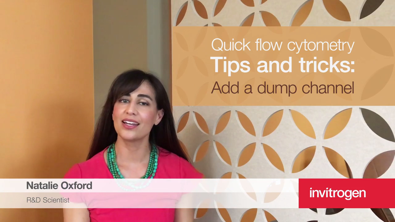
Tip 5: Try to combine negative markers in one channel (dump channel) to save space on your panel
A helpful trick when you want to exclude a lot of cell types at once without having to suck up multiple channels for that would be to use a dump channel. This is where you're placing all the antibodies that identify your cells that are not of interest into the same channel with the same fluorochrome, and then those can be easily gated out and all of the cells negative for the dump channel would be those that you use for your analysis going forward.
Immunology at work
Learn immune cell type markers and protocols. The Invitrogen Immunology at Work Resource Center is a learning center with technical content designed for new and experienced life scientists alike exploring the field of immunology.
Not for resale. Super Bright Polymer Dyes are sold under license from Becton, Dickinson and Company. Brilliant Violet and PE CF dyes are subject to proprietary rights of Becton, Dickinson and Company. Cy™ is a trademark of Amersham Biosciences Corp. Cy dyes are subject to proprietary rights of Amersham Biosciences Corp and Carnegie Mellon University and are made and sold under license from Amersham Biosciences Corp only for research and in vitro diagnostic use. BRILLIANT VIOLET™ is a trademark or registered trademark of Becton, Dickinson and Company or its affiliates, and is used under license. Powered by Sirigen™.