Search Thermo Fisher Scientific
- Contact Us
- Quick Order
-
Don't have an account ? Create Account
Search Thermo Fisher Scientific


| Catalog Number | Volume (Metric) |
|---|---|
| 11161D | 0.4 mL |
| 11131D | 2 mL |
| 11132D | 5 x 2 mL |
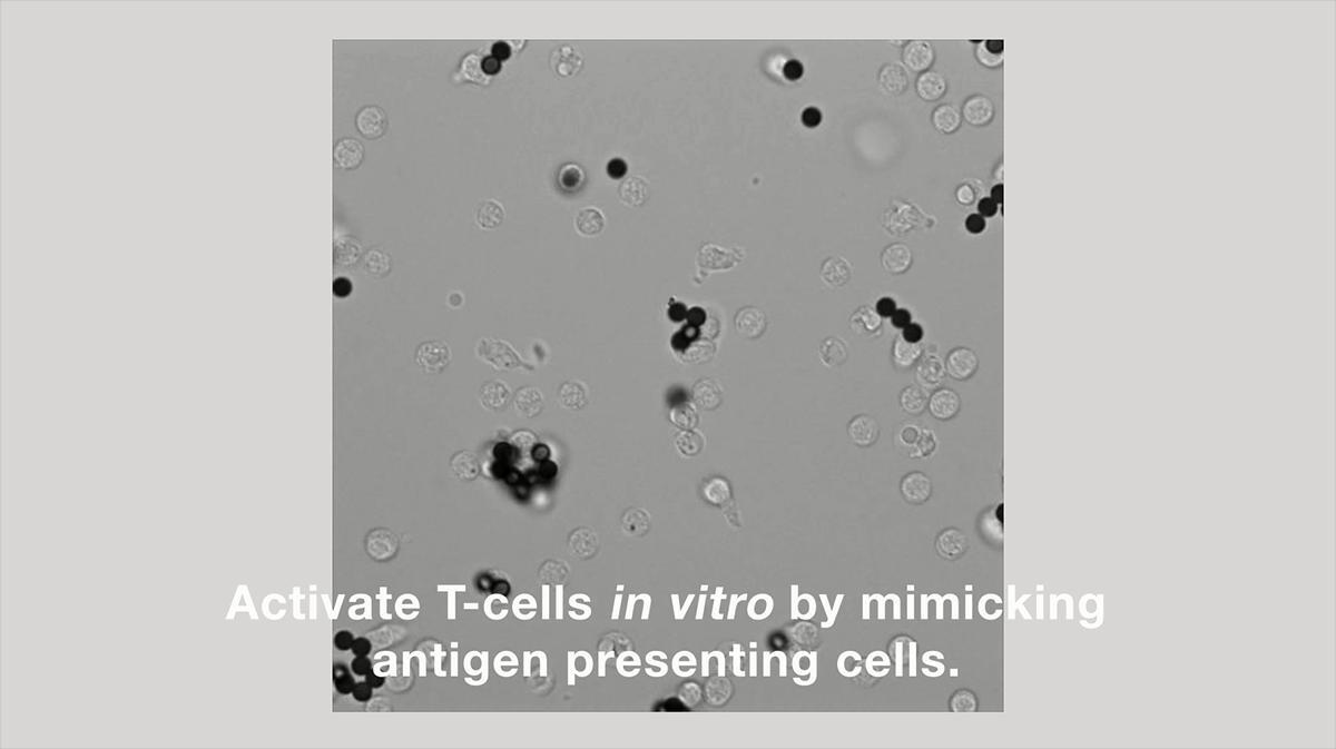
Dynabeads Human T-Activator CD3/CD28 beads are 4.5-μm superparamagnetic beads covalently coupled with an optimized combination of anti-human CD3 and anti-human CD28 antibodies and intended for ex vivo activation and expansion/proliferation of human T cells. T cells are one of the most important cells in the immune system and are responsible for fighting off infections and cancer and are therefore intensively used in many cancer and disease studies.
Benefits of Dynabeads Human T-Activator CD3/CD28 beads include:
• Easy—activate and expand T cells without using feeder cells (antigen-presenting cells)
• Efficient—reproducible method and high T-cell expansion rate (up to 1,000-fold expansion)
• No contamination—using magnetic beads avoids contamination of soluble antibodies or mitogens
• Functional—activated T cells retain in vivo-like function
Simple and fast procedure
Dynabeads Human T-Activator CD3/CD28 beads offer a simple method for the activation and expansion of CD3+ T cells, CD4+ T helper cells, or CD8+ T cytotoxic cells. Pre-isolated T cells are activated by adding the beads and human recombinant IL-2 (rIL-2) to the cell culture. Optimal expansion is performed when appropriate T-cell medium is used, such as CTS OpTmizer T Cell Expansion SFM. Human T cells activated with Dynabeads Human T-Activator CD3/CD28 beads can be used to stimulate B cell proliferation and plasma B cell development.
Activation of human T cells
Short time activation: activation of T cells with Dynabeads Human T-Activator CD3/CD28 beads for a few hours to a few days. This procedure does not expand the cells, and proteomics expression/detection and nucleic acid assays can be performed. The cell:bead complexes cannot be used in flow cytometry in this phase.
Long-term activation/expansion: activation of T cells with Dynabeads Human T-Activator CD3/CD28 beads for three days to a few weeks to obtain an expansion of the T cells. When experiencing reduced expansion or cell shrinking, the T cells can be restimulated with the Dynabeads Human T-Activator CD3/CD28 beads. The beads can be removed with a DynaMag magnet and used in applications like cell:cell interaction studies, flow cytometry, proteomics, or nucleic acid-based assays.
Pre-isolate the human T cells
To obtain the most optimal activation and expansion, pure T cells can be isolated directly from human whole blood or from peripheral blood mononuclear cells (PBMCs) with Dynabeads magnetic beads. For positive bead-based isolation of human CD3, CD4, or CD8 T cells from whole blood or PBMC with a release, use our Dynabeads Human FlowComp products. For negative isolation of human CD3, CD4, or CD8 T cells from PBMC, use our Dynabeads Untouched Human cell products.
Dynabeads Human T-Activator CD3/CD28 applications
The development and execution of chimeric antigen receptor T (CAR-T) cells allow T cells to target specific biomarkers on a range of cell types. CAR-T cells are constructed to have the CD28 and CD3 activating domains on the CAR that greatly enhances the Dynabeads Human T-Activator CD3/CD28 beads. Research into the use specific CAR-T cells against non-small cell lung cancers has demonstrated the importance of the activation beads (Li, et al, 2018). In addition, in vitro activation and expansion of B cells relies entirely on T cell activation. The use of activation beads is paramount for B cell proliferation and differentiation in vitro (Wang et al, 2017).
Alternative activation products, from mouse to clinic
• For ex vivo activation and expansion of human T cells for cell-based therapy, use the aseptically manufactured CTS Dynabeads CD3/CD28
• For activation of mouse T cells, use Dynabeads Mouse T Activator CD3/CD28
• For activation of human regulatory T cells (Tregs), use Dynabeads CD3/CD28 Treg Expander
• For activation of human antigen-specific T cells, use Dynabeads Human T Activator CD3/CD28/CD137
Commercial supply
Our manufacturing sites are ISO 13485–certified. Bead characteristics and manufacturing conditions help ensure consistency and batch reproducibility, making them an ideal choice for commercial supply. If you are utilizing Dynabeads Human T-Activator CD3/CD28 beads in a commercial product or service or need a larger bulk volume of this product, please contact us at oemdynal@lifetech.com or read more on our Dynabeads OEM page.
References
Li N, Liu S, Sun M, Chen W, Xu X, Zeng Z, Tang Y, Dong Y, Chang AH, Zhao Q. Chimeric Antigen Receptor-Modified T Cells Redirected to EphA2 for the Immunotherapy of Non-Small Cell Lung Cancer. Transl Oncol. 2018 Feb;11(1):11-17.
Wang D, Fløisand Y, Myklebust CV, Bürgler S, Parente-Ribes A, Hofgaard PO, Bogen B, Taskén K, Tjønnfjord GE, Schjesvold F, Dalgaard J, Tveita A, Munthe LA. Autologous bone marrow Th cells can support multiple myeloma cell proliferation in vitro and in xenografted mice. Leukemia. 2017 Oct;31(10):2114-2121.


 SDS
SDSPlease review the following possibilities for why your Dynabeads magnetic beads are not pelleting:
- The solution is too viscous.
- The beads have formed aggregates because of protein-protein interaction.
Try these suggestions:
- Increase separation time (leave tub on magnet for 2-5 minutes)
- Add DNase I to the lysate (~0.01 mg/mL)
- Increase the Tween 20 concentration to ~0.05% of the binding and/or washing buffer.
- Add up to 20 mM beta-merecaptoethanol to the binding and/or wash buffers.
Find additional tips, troubleshooting help, and resources within our Dynabeads Nucleic Acid Purification Support Center.
For biotin-labled DNA that is less than 1 kb, we recommend you use Dynabeads M270 Streptavidin and MyOne C1 magnetic beads. We recommend our Dynabeads KilobaseBINDER Kit, which is designed to immobilize long (>1 kb) double-stranded DNA molecules. The KilobaseBINDER reagent consists of M-280 Streptavidin-coupled Dynabeads magnetic beads along with a patented immobilization activator in the binding solution to bind to long, biotinylated DNA molecules for isolation. Please see the following link (https://www.thermofisher.com/us/en/home/life-science/dna-rna-purification-analysis/napamisc/capture-of-biotinylated-targets/immobilisation-of-long-biotinylated-dna-fragments.html) for more information in regards to long biotinylated DNA fragment isolation.
Find additional tips, troubleshooting help, and resources within our Dynabeads Nucleic Acid Purification Support Center.
Yes, Dynabeads magnetic beads can be used to isolate single-stranded DNA. Streptavidin Dynabeads magnetic beads can be used to target biotinylated DNA fragments, followed by denaturation of the double-stranded DNA and removal of the non-biotinylated strand. The streptavidin-coupled Dynabeads magnetic beads will not inhibit any enzymatic activity. This enables further handling and manipulation of the bead-bound DNA directly on the solid phase. Please see the following link (https://www.thermofisher.com/us/en/home/life-science/dna-rna-purification-analysis/napamisc/capture-of-biotinylated-targets/preparing-single-stranded-dna-templates.html) for more information in regards to single-stranded DNA capture.
Find additional tips, troubleshooting help, and resources within our Dynabeads Nucleic Acid Purification Support Center.
Magnetic susceptibility is a measure of how quickly the beads will migrate to the magnet. This will depend on the iron content and the character of the iron oxide. The magnetic susceptibility given for the Dynabeads magnetic beads is the mass susceptibility, given either as cgs units/g or m^3/kg (the latter being an SI unit). For ferri- and ferromagnetic substances, the magnetic mass susceptibility is dependent upon the magnetic field strength (H), as the magnetization of such substances is not a linear function of H but approaches a saturation value with increasing field. For that reason, the magnetic mass susceptibility of the Dynabeads magnetic beads is determined by a standardized procedure under fixed conditions. The magnetic mass susceptibility given in our catalog is thus the SI unit. Conversion from Gaussian (cgs, emu) units into SI units for magnetic mass susceptibility is achieved by multiplying the Gaussian factor (emu/g or cgs/g) by 4 pi x 10^-3. The resulting unit is also called the rationalized magnetic mass susceptibility, which should be distinguished from the (SI) dimensionless magnetic susceptibility unit. In general, magnetic mass susceptibility is a measure of the force (Fz) influencing an object positioned in a nonhomogenous magnetic field. The magnetic mass susceptibility of the Dynabeads magnetic beads is measured by weighing a sample, and then subjecting the sample to a magnetic field of known strength. The weight (F1) is then measured, and compared to the weight of the sample when the magnetic field is turned off (F0). The susceptibility is then calculated as K x 10^-3 = [(F1-F0) x m x 0.335 x 10^6], where K is the mass susceptibility of the sample of mass m. The susceptibility is then converted to SI units.
Find additional tips, troubleshooting help, and resources within our Dynabeads Nucleic Acid Purification Support Center.
There are different methods to check binding of ligands to the beads, including optical density (OD) measurement, fluorescent labeling, and radioactive labeling.
For OD measurement, you would measure the OD of the ligand before immobilization to the beads and compare it with the ligand concentration that is left in the supernatant after coating. This gives a crude measurement of how much protein has bound to the beads.
Protocol:
1.Set spectrophotometer to the right wavelength. As a blank, use the Coupling Buffer.
2.Measure the absorbance of the Pre-Coupling Solution. A further dilution may be necessary to read the absorbance, depending upon the amount of ligand added.
3.Measure the absorbance of the Post-Coupling Solution. A dilution may be necessary to read the absorbance.
4.Calculate the coupling efficiency, expressed as the % protein uptake, as follows. [(Pre-Coupling Solution x D) - (Post-Coupling Solution x D)] x 100/(Pre-Coupling Solution x D) where D = dilution factor.
For fluorescent labeling, we suggest negatively quantifying the amount of ligand bound by measuring ligand remaining in the coupling supernatant (compared to the original sample), rather than directly measuring the ligands on the beads. Add labeled ligand to the beads, and measure how much ligand is left in the supernatant (not bound to the beads). By comparing this with the total amount added in the first place, you can then calculate how much of the ligand that has been bound to the beads. Keep in mind that the Dynabeads magnetic beads are also autofluorescent, which is why direct measuring of fluorescence of the bead-bound ligands is not recommended, but rather this indirect approach. The label could be, for example, FITC/PE. Some researchers perform a direct approach with success (using a flow cytometer).
Radioactive labeling is the most sensitive method of the three, but it is also the most difficult one. It involves radioactively labeling a portion of the ligand. We use radiolabeled I-125 in tracer amounts and mix it with "cold" ligands in a known ratio before coupling. The absolute quantities for the ligand on the beads should be obtained by measuring the beads in a scintillation (gamma) counter and comparing the cpm with a standard.
Protocol:
1.Take out an appropriate amount of beads and wash the beads in 1 mL of binding buffer.
2.Pipette out desired amount of human IgG in a separate tube.
3.Mix the human IgG with I-125-labeled human IgG (30,000 - 100,000 cpm).
4.Dilute the mixture of human IgG and I-125-labeled human IgG to 100 mL in binding buffer.
5.Incubate for 30 minutes at room temperature and measure the cpm in a scintillation counter.
6.Wash the beads (with coating) four times, and measure cpm again.
The % binding is calculated by using the equation : (cpm after washing/cpm before washing)x100%.
Find additional tips, troubleshooting help, and resources within our Dynabeads Nucleic Acid Purification Support Center.
Share catalog number, name or link