Search Thermo Fisher Scientific


Certificates

SDS

Citations & References (68)
Invitrogen™
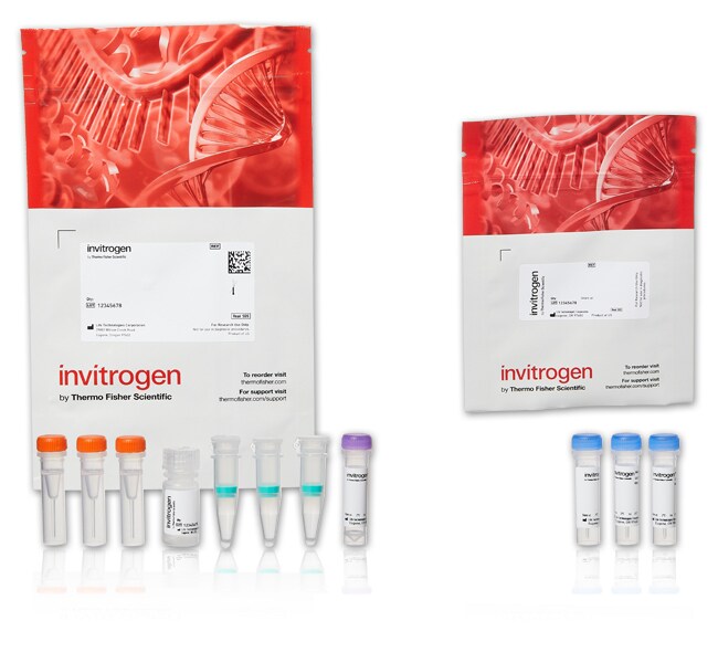
Alexa Fluor™ 488 Microscale Protein Labeling Kit
Microscale Protein Labeling Kits provide a convenient means for attaching a fluorescent label to a small amount of antibody orRead more
 Promotion
Promo code:RPUZZ25
Stock up on essentials to piece your discovery together

Until June 27, save up to $650 and get an exclusive lab-themed hidden-object puzzleLearn More
| Catalog Number | Quantity |
|---|---|
| A30006 | 1 Kit |
Catalog number A30006
Price (USD)
786.00
Each
Estimated availability date 25-Aug-2025
Quantity:
1 Kit
Price (USD)
786.00
Each
Microscale Protein Labeling Kits provide a convenient means for attaching a fluorescent label to a small amount of antibody or protein (20–100 μg). The kits are available in four Alexa Fluor™ colors (or biotin) and supply everything needed for three labeling and separation reactions.
Important Features of Microscale Protein Labeling Kits:
- Labeled proteins typically ready to use typically in 2 hr (∼30 min hands-on time)
- Optimized for 20–100 μg of protein with molecular weights between 12 and 150 kDa
- Purified using convenient spin filters with yields between 60 and 90%
- Stabilizing proteins must be removed from the sample before labeling
Stable Reaction Chemistry and Superior Alexa Fluor™ Dyes
In the Microscale Protein Labeling Kits, the reactive dye contains a tetrafluorophenyl (TFP) ester moiety that is more stable in solution than the commonly used succinimidyl (NHS) ester. TFP esters react efficiently with primary amines of proteins to form stable dye–protein conjugates. Compared to traditional dyes, Alexa Fluor™ dyes are brighter, more photostable, and more pH resistant between pH 4 and 10. And generally when using Alexa Fluor™ dyes, higher degrees of labeling can be achieved without intramolecular quenching. For details see Alexa Fluor™ Dyes Spanning the Visible and Infrared Spectrum—Section 1.3.
Learn More About Protein and Antibody Labeling
We offer a wide selection of Molecular Probes™ antibody and protein labeling kits to fit your starting material and your experimental setup. See Antibody Labeling from A to Z or use our Labeling Chemistry Selection Tool for other choices. To learn more about our various kits read Kits for Labeling Proteins and Nucleic Acids—Section 1.2 in the Molecular Probes™ Handbook.
We'll Make a Custom Antibody Conjugate for You
If you can't find what you're looking for in our stocked list, we'll prepare a custom antibody conjugate for you. Our custom conjugation service is efficient and confidential, and we stand by the quality of our work. We are ISO 13485:2000 certified.
For Research Use Only. Not for use in diagnostic procedures.
Specifications
ColorGreen
Detection MethodFluorescence
Excitation/Emission495/519 nm
Label TypeAlexa Fluor
Labeling MethodConjugation-based
Labeling Scale20–100 μg
Product LineAlexa Fluor™
Product TypeLabeling Kit
Quantity1 Kit
Reactive MoietyTetrafluorophenyl (TFP) Ester
Shipping ConditionRoom Temperature
Labeling TargetProteins
Label or DyeAlexa Fluor 488
Unit SizeEach
Contents & Storage
Store in refrigerator 2°C to 8°C and protect from light.

Have questions about this product? Ask our AI assisted search.
What formulation of antibody should I use for conjugation for small animal in vivo imaging?
Can I use 50 μg of protein with Fluorescent Protein Labeling Kits?
What is degree of labeling (DOL)?
This is an AI-powered search and may not always get things right. You can help us make it better with a thumbs up or down on individual answers or by selecting the “Give feedback" button. Your search history and customer login information may be retained by Thermo Fisher and processed in accordance with our
Privacy Notice.
Figures

Chemical Structure
Customers who viewed this item also viewed
Documents & Downloads
Certificates
Search by lot number or partial lot number
Lot #Certificate TypeDateCatalog Number(s)
3086991Certificate of AnalysisDec 10, 2024A30006
2770809Certificate of AnalysisDec 22, 2023A30006
2413458Certificate of AnalysisDec 01, 2023A30006
2626258Certificate of AnalysisJul 05, 2023A30006
2381714Certificate of AnalysisSep 23, 2021A30006
5 results displayed, search above for a specific certificate
Safety Data Sheets
SDSProduct Information
Frequently asked questions (FAQs)
No. We recommend using 1 mg of protein with Fluorescent Protein Labeling Kits. For smaller protein sample sizes, we recommend using Microscale Protein Labeling kits which are optimized for 20-100 µg of protein.
Find additional tips, troubleshooting help, and resources within our Cell Analysis Support Center.
To allow for good reaction kinetics, antibodies should be in PBS buffer at a concentration of 0.5-3.0 mg/ml. The antibody must be free of preservatives (azide etc.), amine containing buffers and carrier proteins such as BSA.
Find additional tips, troubleshooting help, and resources within our Cell Analysis Support Center.
Degree of labeling (DOL) describes the number of fluorophores per antibody. For in vivo labeling experiments, the DOL is restricted to a narrow range because it has significant consequences for the biodistribution and clearance of the probe. For example, for in vivo imaging, we have determined that the DOL range for the far-red Alexa Fluor dyes is 1.5 to 3 molecules per antibody for optimal optical in vivo imaging.
Find additional tips, troubleshooting help, and resources within our Cell Analysis Support Center.
Citations & References (68)
Search citations by name, author, journal title or abstract text
Citations & References
Abstract
Golgi apparatus immunolocalization of endomannosidase suggests post-endoplasmic reticulum glucose trimming: implications for quality control.
Journal:Mol Biol Cell
PubMed ID:11102520
'Trimming of N-linked oligosaccharides by endoplasmic reticulum (ER) glucosidase II is implicated in quality control of protein folding. An alternate glucosidase II-independent deglucosylation pathway exists, in which endo-alpha-mannosidase cleaves internally the glucose-substituted mannose residue of oligosaccharides. By immunogold labeling, we detected most endomannosidase in cis/medial Golgi cisternae (83.8% of immunogold
Glycosylation influences the lectin activities of the macrophage mannose receptor.
Journal:J Biol Chem
PubMed ID:15983039
'The mannose receptor (MR) is a heavily glycosylated endocytic receptor that recognizes both mannosylated and sulfated ligands through its C-type lectin domains and cysteine-rich (CR) domain, respectively. Differential binding properties have been described for MR isolated from different sources, and we hypothesized that this could be due to altered glycosylation.
An endocytosed TGN38 chimeric protein is delivered to the TGN after trafficking through the endocytic recycling compartment in CHO cells.
Journal:J Cell Biol
PubMed ID:9722606
'To examine TGN38 trafficking from the cell surface to the TGN, CHO cells were stably transfected with a chimeric transmembrane protein, TacTGN38. We used fluorescent and 125I-labeled anti-Tac IgG and Fab fragments to follow TacTGN38''s postendocytic trafficking. At steady-state, anti-Tac was mainly in the TGN, but shortly after endocytosis it
Alexa dyes, a series of new fluorescent dyes that yield exceptionally bright, photostable conjugates.
Journal:J Histochem Cytochem
PubMed ID:10449539
'Alexa 350, Alexa 430, Alexa 488, Alexa 532, Alexa 546, Alexa 568, and Alexa 594 dyes are a new series of fluorescent dyes with emission/excitation spectra similar to those of AMCA, Lucifer Yellow, fluorescein, rhodamine 6G, tetramethylrhodamine or Cy3, lissamine rhodamine B, and Texas Red, respectively (the numbers in the
Removal of the membrane-anchoring domain of epidermal growth factor leads to intracrine signaling and disruption of mammary epithelial cell organization.
Journal:J Cell Biol
PubMed ID:9832559
'Autocrine EGF-receptor (EGFR) ligands are normally made as membrane-anchored precursors that are proteolytically processed to yield mature, soluble peptides. To explore the function of the membrane-anchoring domain of EGF, we expressed artificial EGF genes either with or without this structure in human mammary epithelial cells (HMEC). These cells require activation
68 total citations