Search Thermo Fisher Scientific

MitoTracker™ Dyes for Mitochondria Labeling
| 產品號碼 | Description | Excitation Wavelength Range |
|---|---|---|
| M22426 | MitoTracker Deep Red FM | 644/665 nm |
| M22425 | MitoTracker™ Red FM | 581/644 nm |
| M7512 | MitoTracker™ Red CMXRos | 579/599 nm |
| M7513 | MitoTracker Red CM-H2Xros | 579/599 nm |
| M7514 | MitoTracker™ Green FM | 490/516 nm |
| M7510 | MitoTracker Orange CMTMRos | 554/576 nm |
| M7511 | MitoTracker Orange CM-H2TMRos | 554/576 nm |
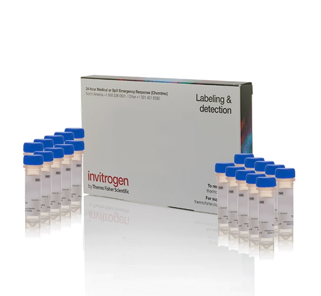
Rosamine & carbocyanine-based staining dyes MitoTracker Orange CMTMRos, Red CMXRos, Orange CM-H2TMRos, Red CM-H2XRos, Red FM, Green FM, & Deep Red FM enable mitochondria visualization with fluorescent imaging. These mitochondria dyes are specially packaged in vials of 50 μg each to create freshly reconstituted dyes without the impact of freeze-thaw cycles to aid in a simplified sample prep for cell analysis workflow.
Rosamine-based MitoTracker dyes for mitochondria labeling
Cell-permeant MitoTracker probes stain active mitochondria in live cells for labeling and localization in fluorescent cell imaging. Our rosamine-based mitochondrial staining dyes include MitoTracker Orange CMTMRos, a derivative of tetramethylrosamine, and MitoTracker Red CMXRos, a derivative of X-rosamine. MitoTracker Orange CM-H2TMRos and MitoTracker Red CM-H2XRos are reduced, nonfluorescent versions of the rosamine-based MitoTracker dyes that fluoresce upon oxidation. The fluorescent signal from the rosamine-based MitoTracker dyes is retained in the mitochondria even after aldehyde fixation and detergent permeabilization, making these mitochondria dyes flexible for many workflows including applications that require subsequent processing such as immunocytochemistry or in situ hybridization. MitoTrackerRed and Orange are well suited for multicolor labeling experiments because their fluorescence is well resolved from the green fluorescence of other probes.
Carbocyanine-based MitoTracker dyes for mitochondria labeling
Cell-permeant MitoTracker dyes stain active mitochondria in live cells for mitochondrial labeling and localization in fluorescent cell imaging. The carbocyanine-based dyes MitoTracker Green FM and MitoTracker Red FM accumulate in active mitochondria but are not well-retained in mitochondria after aldehyde fixation. The carbocyanine-based MitoTracker Deep Red FM stains active mitochondrial in live cells and is well-retained in mitochondria after aldehyde fixation and subsequent permeabilization with detergents for applications that require subsequent processing such as immunocytochemistry or in situ hybridization. MitoTracker Red FM and MitoTracker Deep Red FM are well suited for multicolor labeling experiments because their red fluorescence is well resolved from the green fluorescence of other probes.
Benefits of using MitoTracker mitochondria labeling dyes
MitoTracker dyes are provided in vials of 50 μg of lyophilized powder ready for reconstitution. To label mitochondria, live cells are simply incubated with the MitoTracker probe of your choice. The mitochondrial staining dyes passively diffuse across the plasma membrane and accumulate in active mitochondria. The MitoTracker dyes are offered in a range of wavelengths and can be used for mitochondrial localization in multicolor experiments.
Conventional fluorescent stains such as tetramethylrosamine and rhodamine 123 are readily sequestered by active mitochondria and are reversible in dynamic membrane potential measurements as they easily wash out of cells upon loss in membrane potential. In contrast, MitoTracker dyes contain a mildly thiol-reactive chloromethyl moiety so that mitochondrial staining is retained if the mitochondrial membrane potential is lost, allowing many of the MitoTracker dyes to be retained during cell fixation.
Throughout the cell life cycle, mitochondria use oxidizable substrates to produce an electrochemical proton gradient across the mitochondrial membrane (whose potential is negative), resulting in ATP production. MitoTracker dyes are ideal probes for mitochondria staining in experiments studying the cell cycle or processes such as apoptosis and other end point assays. MitoTracker dyes are also available for flow cytometry applications (Cat. No. M46750, M46751, M46752, and M46753).
Related products for mitochondrial membrane potential
For studying dynamic mitochondria membrane potential specifically, we recommend using JC-1 (cationic carbocyanine dye, Cat. No. T3168) or TMRM (tetramethyl rhodamine methyl ester, Cat. No. I34361) dyes. The MitoProbe JC-1 Assay Kit (Cat. No. M34152) contains the JC-1 dye in addition to the potent mitochondrial membrane-potential disrupter, CCCP, which depolarizes the mitochondrial membrane. These reagents can provide compensatory controls to correctly compensate green-to-red fluorescence ratio. TMRM is used for the detection of dynamic measurement of mitochondrial membrane potential.
圖表


瀏覽此商品的人,也瀏覽
文件與下載
證書
安全資料表
Scientific Resources
Product Information
常見問答集 (常見問題)
This is typically a result of using too high of a concentration of the Mitotracker dye. Most organic dyes are used in the low micromolar range. The MitoTracker dyes are used at a much lower concentration, around 50–200 nanomolar. Higher concentrations can cause background fluorescence and non-mitochondrial staining.
Find additional tips, troubleshooting help, and resources within our Cell Analysis Support Center.
Yes, it has been done. A literature search will find numerous examples. However, be aware that plate readers are typically less sensitive than microscopes or flow cytometers, which means that the degree of change in fluorescence may not be as sensitive or easily detectable with a plate reader. Be sure to optimize label times and concentrations carefully.
Find additional tips, troubleshooting help, and resources within our Cell Analysis Support Center.
Yes, this is possible because MitoTracker Deep Red FM dye is retained upon fixation. See Table 2 in this manual (https://assets.thermofisher.com/TFS-Assets/LSG/manuals/mp07510.pdf).
Find additional tips, troubleshooting help, and resources within our Cell Analysis Support Center.
The Mitotracker dyes should be imaged soon after staining because over time, those dyes can be toxic.
Find additional tips, troubleshooting help, and resources within our Cell Analysis Support Center.
Regardless of which dye you use - tetramethylrhodamine, methyl ester (TMRM), JC-1 or MitoTracker - untreated cells will fluoresce. It's just that cells with reduced mitochondrial membrane potential will fluoresce less. It is the degree of change which is important. JC-1 dye not only changes intensity, but has a ratiometric spectral change in excitation and emission. It is very important to have an untreated control as well as a positive control treated with a mitochondrial membrane potential destabilizer, such as CCCP or FCCP. Most mitochondrial stains are only for use with live cells, as the signal will not be retained to the same degree with fixation.
Find additional tips, troubleshooting help, and resources within our Cell Analysis Support Center.
