Search Thermo Fisher Scientific

c7c8 - Key/Specs/Resources/Appl/Tech/Doc/Contact
Field emission gun scanning electron microscope
The Thermo Scientific Phenom Pharos G2 FEG-SEM brings field emission SEM to your tabletop. The Phenom Pharos G2 FEG-SEM will outperform many floor-standing SEMs in terms of image quality, while offering a vastly better user experience. For academic and industrial laboratories that so far did not consider SEM a realistic option, the Phenom Pharos G2 FEG-SEM makes FEG performance accessible thanks to its attractive form factor and short training required. Blazing fast sample loading means fast sample exchange, which means higher productivity. Unlike other SEMs, which end up being fully booked, the Phenom Pharos G2 FEG-SEM performs imaging and analysis jobs so quickly that it serves well as a walk-up tool.
The new Phenom Pharos G2 FEG-SEM expands its acceleration voltage range down to 1 kV, to better accommodate insulating and beam-sensitive samples, and up to 20 kV, with a resolution of 2.0 nm that reveals the finest details.
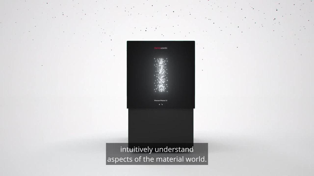
Introducing the Phenom Pharos G2 Desktop FEG-SEM: Intuitive evidence for your research
Unique field emission source
Unique among desktop SEMs, the Phenom Pharos G2 FEG-SEM offers a field emission source, which guarantees high brightness, crisp images, and stable beam current.
Excellent resolving power
The Phenom Pharos G2 FEG-SEM offers a resolution of 2.0 nm at 20 kV. Such performance shows the shape of nanoparticles, imperfections in coatings, or other features that would be missed by tungsten SEMs or other tabletop SEMs.
Gentle imaging
With a voltage range down to 1 kV, the Phenom Pharos G2 FEG-SEM enables imaging of beam-sensitive samples, such as polymers, as well as insulating samples, without the requirement to apply a coating. As a result, nanoscale surface features are not obscured.
Higher productivity
While FEG SEMs have a reputation for being difficult to accommodate and difficult to operate, the Phenom Pharos G2 FEG-SEM literally requires only a desk, and less than one hour of training. Master students, visitors, or other researchers typically not trained to work on high-end FEG SEMs can easily use the Phenom Pharos G2 FEG-SEM to create eye-catching images.
A world of information
On the Phenom Pharos G2 FEG-SEM, morphological information is acquired together with compositional information, thanks to SE, BSE, and EDS detectors built into the system. A range of sample holders is available for temperature-controlled or electrical experiments.

ChemiSEM Technology
Thermo Scientific ChemiSEM Technology revolutionizes and simplifies EDS analysis by fully integrating SEM and EDS functions into a single, cohesive user interface. Based on live quantification and building on decades of expertise in EDS analysis, the technology provides elemental information quickly and easily, guaranteeing reliable results in less time. ChemiSEM Technology now comes with a powerful new feature: ChemiPhase. ChemiPhase identifies unique phases with a big data approach, finding minor and trace elements while eliminating user bias and reducing possible mistakes.
Style Sheet for Products Table Specifications
| Resolution |
|
| Electron optical magnification range |
|
| Light optical magnification |
|
| Acceleration voltages |
|
| Vacuum modes |
|
| Detector |
|
| Sample size |
|
| Sample height |
|
Style Sheet for Komodo Tabs
, significant charging artefacts can still be observed. At 1 kV (right image), those artifacts have disappeared.")
Uncoated insulators, such as this sea shell, can only be properly imaged at low acceleration voltage. At 2 kV (left image), significant charging artifacts can still be observed. At 1 kV (right image), those artifacts have disappeared.

Sensitive materials require gentle conditions. With an acceleration voltage down to 1 kV, the Phenom Pharos G2 Desktop FEG-SEM images beam-sensitive samples without sample coating or other sample preparation. Left: pharmaceutical powder, imaged without damage at 1 kV. Right: the same sample imaged at 5 kV, with damage, illustrating the need for low-kV imaging.

Silver nanostructure

Gold nanoparticles
")
CaCO3 powder (Coated)

Coated Polymer Film

Lung tissue

Phenom User Interface

EDS & Live EDS
 Pharos G2 Desktop Introduction
Introducing the Phenom Pharos G2 Desktop FEG-SEM: Intuitive evidence for your research
 Pharos G2 demo video
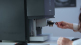
This video will show you how easy a high-performance field emission SEM can be. The Phenom Pharos G2 desktop FEG-SEM only requires a short training in order to run imaging and analysis jobs, and it is very fast.
 Phenom World - Pharos introduction movie
Phenom Pharos
 Phenom Pharos SEM Launch - David Wall
Phenom Pharos SEM Launch - David Wall
 Demo Impression - Mark Elliot
Pharos Demo Impression - Mark Elliot
 Demo Impression - Alex Tamok
Pharos Demo Impression - Alex Tamok
 Demo Impression - Khalid Rana
Pharos Demo Impression - Khalid Rana
 Phenom Pharos SEM Launch - Jos Maas
Phenom Pharos SEM Launch - Jos Maas
 Demo Impression - Christopher Smart
Demo Impression - Christopher Smart
 Pharos Demo Impression - Researchers, Mexico
Pharos Demo Impression - Researchers, Mexico
Webinar: Scanning electron microscopy: selecting the right technology for your needs
This on-demand webinar has been designed to help you decide which SEM best meets your unique needs. We present an overview of Thermo Fisher Scientific SEM technology for multi-user research labs and focus on how these wide-ranging solutions deliver performance, versatility, in situ dynamics and faster time to results. Watch this webinar if you are interested in:
- How the needs for different microanalysis modalities are met (EDX, EBSD, WDS, CL, etc.).
- How samples are characterized in their natural state without the need for sample preparation.
- How new advanced automation allows researchers to save time and increase productivity.
Uncoated insulators, such as this sea shell, can only be properly imaged at low acceleration voltage. At 2 kV (left image), significant charging artifacts can still be observed. At 1 kV (right image), those artifacts have disappeared.
Sensitive materials require gentle conditions. With an acceleration voltage down to 1 kV, the Phenom Pharos G2 Desktop FEG-SEM images beam-sensitive samples without sample coating or other sample preparation. Left: pharmaceutical powder, imaged without damage at 1 kV. Right: the same sample imaged at 5 kV, with damage, illustrating the need for low-kV imaging.
Silver nanostructure
Gold nanoparticles
CaCO3 powder (Coated)
Coated Polymer Film
Lung tissue
Phenom User Interface
EDS & Live EDS
- Pharos G2 Desktop Introduction
Introducing the Phenom Pharos G2 Desktop FEG-SEM: Intuitive evidence for your research
- Pharos G2 demo video
This video will show you how easy a high-performance field emission SEM can be. The Phenom Pharos G2 desktop FEG-SEM only requires a short training in order to run imaging and analysis jobs, and it is very fast.
- Phenom World - Pharos introduction movie
Phenom Pharos
- Phenom Pharos SEM Launch - David Wall
Phenom Pharos SEM Launch - David Wall
- Demo Impression - Mark Elliot
Pharos Demo Impression - Mark Elliot
- Demo Impression - Alex Tamok
Pharos Demo Impression - Alex Tamok
- Demo Impression - Khalid Rana
Pharos Demo Impression - Khalid Rana
- Phenom Pharos SEM Launch - Jos Maas
Phenom Pharos SEM Launch - Jos Maas
- Demo Impression - Christopher Smart
Demo Impression - Christopher Smart
- Pharos Demo Impression - Researchers, Mexico
Pharos Demo Impression - Researchers, Mexico
Webinar: Scanning electron microscopy: selecting the right technology for your needs
This on-demand webinar has been designed to help you decide which SEM best meets your unique needs. We present an overview of Thermo Fisher Scientific SEM technology for multi-user research labs and focus on how these wide-ranging solutions deliver performance, versatility, in situ dynamics and faster time to results. Watch this webinar if you are interested in:
- How the needs for different microanalysis modalities are met (EDX, EBSD, WDS, CL, etc.).
- How samples are characterized in their natural state without the need for sample preparation.
- How new advanced automation allows researchers to save time and increase productivity.

Process control using electron microscopy
Modern industry demands high throughput with superior quality, a balance that is maintained through robust process control. SEM and TEM tools with dedicated automation software provide rapid, multi-scale information for process monitoring and improvement.

Quality control and failure analysis
Quality control and assurance are essential in modern industry. We offer a range of EM and spectroscopy tools for multi-scale and multi-modal analysis of defects, allowing you to make reliable and informed decisions for process control and improvement.

Fundamental Materials Research
Novel materials are investigated at increasingly smaller scales for maximum control of their physical and chemical properties. Electron microscopy provides researchers with key insight into a wide variety of material characteristics at the micro- to nano-scale.

Energy Dispersive Spectroscopy
Energy dispersive spectroscopy (EDS) collects detailed elemental information along with electron microscopy images, providing critical compositional context for EM observations. With EDS, chemical composition can be determined from quick, holistic surface scans down to individual atoms.
_Technique_800x375_144DPI.jpg)
EDS Elemental Analysis
Thermo Scientific Phenom Elemental Mapping Software provides fast and reliable information on the distribution of chemical elements within a sample.
_Technique_800x375_144DPI.jpg)
3D EDS Tomography
Modern materials research is increasingly reliant on nanoscale analysis in three dimensions. 3D characterization, including compositional data for full chemical and structural context, is possible with 3D EM and energy dispersive X-ray spectroscopy.

Atomic-Scale Elemental Mapping with EDS
Atomic-resolution EDS provides unparalleled chemical context for materials analysis by differentiating the elemental identity of individual atoms. When combined with high-resolution TEM, it is possible to observe the precise organization of atoms in a sample.

Imaging Hot Samples
Studying materials in real-world conditions often involves working at high temperatures. The behavior of materials as they recrystallize, melt, deform, or react in the presence of heat can be studied in situ with scanning electron microscopy or DualBeam tools.

In Situ experimentation
Direct, real-time observation of microstructural changes with electron microscopy is necessary to understand the underlying principles of dynamic processes such as recrystallization, grain growth, and phase transformation during heating, cooling, and wetting.

Multi-scale analysis
Novel materials must be analyzed at ever higher resolution while retaining the larger context of the sample. Multi-scale analysis allows for the correlation of various imaging tools and modalities such as X-ray microCT, DualBeam, Laser PFIB, SEM and TEM.
Energy Dispersive Spectroscopy
Energy dispersive spectroscopy (EDS) collects detailed elemental information along with electron microscopy images, providing critical compositional context for EM observations. With EDS, chemical composition can be determined from quick, holistic surface scans down to individual atoms.
EDS Elemental Analysis
Thermo Scientific Phenom Elemental Mapping Software provides fast and reliable information on the distribution of chemical elements within a sample.
3D EDS Tomography
Modern materials research is increasingly reliant on nanoscale analysis in three dimensions. 3D characterization, including compositional data for full chemical and structural context, is possible with 3D EM and energy dispersive X-ray spectroscopy.
Atomic-Scale Elemental Mapping with EDS
Atomic-resolution EDS provides unparalleled chemical context for materials analysis by differentiating the elemental identity of individual atoms. When combined with high-resolution TEM, it is possible to observe the precise organization of atoms in a sample.
Imaging Hot Samples
Studying materials in real-world conditions often involves working at high temperatures. The behavior of materials as they recrystallize, melt, deform, or react in the presence of heat can be studied in situ with scanning electron microscopy or DualBeam tools.
In Situ experimentation
Direct, real-time observation of microstructural changes with electron microscopy is necessary to understand the underlying principles of dynamic processes such as recrystallization, grain growth, and phase transformation during heating, cooling, and wetting.
Multi-scale analysis
Novel materials must be analyzed at ever higher resolution while retaining the larger context of the sample. Multi-scale analysis allows for the correlation of various imaging tools and modalities such as X-ray microCT, DualBeam, Laser PFIB, SEM and TEM.
Style Sheet to change H2 style to p with em-h2-header class
Electron microscopy services for
the materials science
To ensure optimal system performance, we provide you access to a world-class network of field service experts, technical support, and certified spare parts.
Electron microscopy support and resources
Style Sheet for Support and Service footer
Style Sheet for Fonts
Style Sheet for Cards