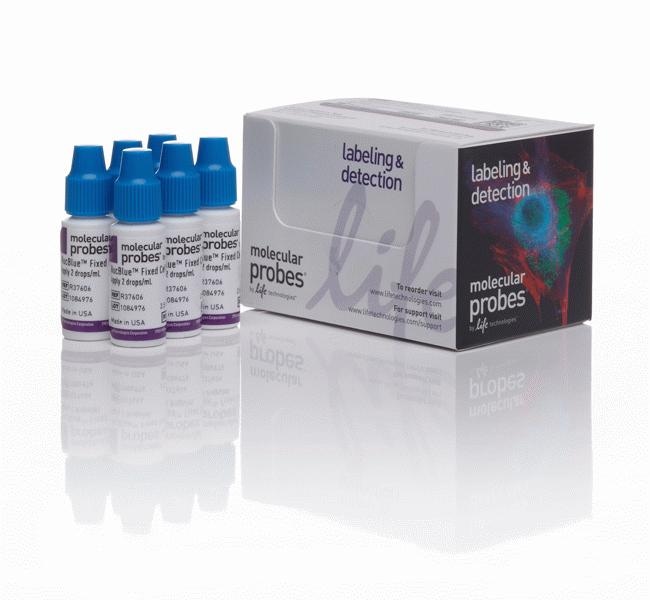
NucBlue™ Fixed Cell ReadyProbes™ Reagent (DAPI)
Invitrogen™

NucBlue™ Fixed Cell ReadyProbes™ Reagent (DAPI)

DAPI is a commonly used nuclear counterstain for fixed cells that emits blue fluorescence when bound to DNA. With NucBlueRead more
Have Questions?
Catalog NumberQuantity
R376066 vial(s) kit
Catalog number R37606
Price (USD)
194.00
Each
In stock
Add to cart
Quantity:
6 vial(s) kit
Recurring order eligible. Learn more »
Price (USD)
194.00
Each
Add to cart
Ask our AI about this Product
DAPI is a commonly used nuclear counterstain for fixed cells that emits blue fluorescence when bound to DNA. With NucBlue Fixed Cell ReadyProbes Reagent, we have formulated a high-purity form of this classic stain in a room temperature-stable solution that is provided in a convenient-to-use dropper bottle. Just tip and drip two drops per ml to stain your cells.

Also available: DAPI (4',6-diamidino-2-phenylindole, dihydrochloride) at 12.5X concentration (5 μM) in water.

• No need to dilute, weigh, or pipette
• Convenient dropper bottle—just use two drops per mL
• Stable at room temperature—keep handy at your scope or cell culture area• DAPI is excited with UV light and detected through a blue/cyan filter

See other ReadyProbes reagents for cell staining
See other nuclear stains for imaging

Cell imaging applications
DAPI is a classic fluorescent dye used extensively for nuclear staining of fixed cells. In fluorescence microscopy, DAPI is excited with UV light and detected through a blue/cyan filter (Figure 1)..

Suggestions for use
In most cases 2 drops/ml of NucBlue Fixed Cell Stain and an incubation of 15 to 30 minutes will produce bright nuclear staining; however, optimization may be needed for some cell types, conditions, and applications. In such cases, simply add more, or fewer, drops until the optimal staining intensity is obtained.
• NucBlue Fixed Cell Stain is excited by UV light at 360 nm when bound to DNA, with an emission maximum at 460 nm. It is detected through a blue/cyan filter, such as a DAPI filter, blue GFP filters, or the Semrock BrightLine Alexa Fluor 350 Dye filter set.
• As a preferred blue nuclear stain in fixed cell imaging experiments, NucBlue Fixed Cell Stain is ideal for use with antibody-based applications.

Visualize staining your cell without wasting your reagents, antibodies, or time with our new Stain-iT Cell Staining Simulator.

For Research Use Only. Not for use in diagnostic procedures.
Specifications
ColorBlue
Detection MethodFluorescence
Dye TypeDAPI
EmissionVisible
Excitation Wavelength Range360⁄460
For Use With (Equipment)Fluorescence Microscope, Flow Cytometer
FormLiquid
Product LineMolecular Probes™
Quantity6 vial(s) kit
Shipping ConditionRoom Temperature
Label TypeFluorescent Dye
Product TypeNucleic Acid Stain
SubCellular LocalizationNucleus
Unit SizeEach
Contents & Storage
6 × 2.5 mL dropper bottles

Store at ≤ 25°C

Have questions about this product? Ask our AI assisted search.
Can I use the ReadyProbes reagents for flow cytometry?
Do you offer an alternative for the discontinued 3-Germ Layer ICC Kit (Cat. No. A25538)?
My DAPI labeled samples have a strong blue background signal immediately after mounting. What can I do to fix this?
This is an AI-powered search and may not always get things right. You can help us make it better with a thumbs up or down on individual answers or by selecting the “Give feedback" button. Your search history and customer login information may be retained by Thermo Fisher and processed in accordance with our Privacy Notice.

Figures

Videos

EVOS M7000 microscope Z-stack of rat cortical neurons using a 20X S-Apo objective, DAPI, GFP and RFP LED cubes.

Video Player is loading.
Current Time 0:00
Duration 0:00
Loaded: 0%
Stream Type LIVE
Remaining Time 0:00
 
1x
    • Chapters
    • descriptions off, selected
    • captions off, selected

      Rat cortical neurons (A1084001) transfected with CellLight??? Golgi-RFP, BacMam 2.0 (C10593). At 48 hours cells were fixed and stained with anti-Tau (MN1000), followed by Goat anti-Mouse Alexa Fluor Plus 488 (A32723) and NucBlue??? Fixed Cell ReadyProbes??? Reagent (R37606). Cells were imaged on an M7000 (AMF7000) using an Olympus 20X objective (AMEP4734) with DAPI (AMEP4650), GFP (AMEP4651) and RFP (AMEP4652) LED cubes.

      Customers who viewed this item also viewed


      Documents & Downloads

      Certificates

      Lot #Certificate TypeDateCatalog Number(s)
      3123552Certificate of AnalysisFeb 09, 2025R37606
      3001522Certificate of AnalysisAug 14, 2024R37606
      2911366Certificate of AnalysisApr 17, 2024R37606
      2831256Certificate of AnalysisFeb 27, 2024R37606
      2747486Certificate of AnalysisNov 02, 2023R37606
      5 results displayed, search above for a specific certificate

      Safety Data Sheets

      Frequently asked questions (FAQs)

      Some mounting medias can have strong blue autofluorescence. If you are seeing a high blue background, it could be coming from the mountant. Try labeling the sample and view it before (using a wet mount in buffer) and after mounting to determine if the background signal is coming from the mounting media or the sample itself.

      Find additional tips, troubleshooting help, and resources within our Cell Analysis Support Center.

      We do not have a direct alternative for the discontinued 3-Germ Layer ICC Kit (Cat. No. A25538). However, we do have alternative primary and secondary antibodies, as well as reagents, that can be used for trilineage differentiation. Please note that we have not internally validated the use of all these reagents together.

      We recommend the following primary antibodies:

      Alpha-Smooth Muscle Actin Monoclonal Antibody (1A4 (asm-1)) (Cat. No. MA5-11547)

      alpha-Fetoprotein Monoclonal Antibody (AFP3) (Cat. No. 14-6583-80)

      beta-3 Tubulin Monoclonal Antibody (2G10) (Cat. No. MA1-118)

      We recommend the following secondary antibodies:

      Goat anti-Mouse IgG2a Cross-Adsorbed, Alexa Fluor 555 (Cat. No. A21137)

      Goat anti-Mouse IgG2a Cross-Adsorbed, Alexa Fluor 594 (Cat. No. A21135)

      https://www.thermofisher.com/antibody/product/Goat-anti-Mouse-IgG1-Cross-Adsorbed-Secondary-Antibody-Polyclonal/A-21121

      We recommend using the following reagents:

      https://www.thermofisher.com/order/catalog/product/88-8824-00

      NucBlue Fixed Cell ReadyProbes Reagent (DAPI) (Cat. No. R37606)

      Blocker BSA (Cat. No. 37520)

      DPBS (10X), no calcium, no magnesium (Cat. No. 14200075)

      Find additional tips, troubleshooting help, and resources within our Cell Analysis Support Center.

      This is not recommended. The ReadyProbes reagents were developed for imaging applications whereas the Ready Flow reagents were optimized for flow cytometry.

      Find additional tips, troubleshooting help, and resources within our Cell Analysis Support Center.

      Citations & References (29)

      Citations & References
      Abstract
      Multipotent stem cells from trabecular meshwork become phagocytic TM cells.
      Authors:Du Y, Roh DS, Mann MM, Funderburgh ML, Funderburgh JL, Schuman JS,
      Journal:Invest Ophthalmol Vis Sci
      PubMed ID:22297497
      'To isolate and characterize stem cells from human trabecular meshwork (TM) and to investigate the potential of these stem cells to differentiate into TM cells. Human trabecular meshwork stem cells (TMSCs) were isolated as side population cells by fluorescence-activated cell sorting or isolated by clonal cultures. Passaged TMSCs were compared ... More
      Transitions of protein traffic from cardiac ER to junctional SR.
      Authors:Sleiman NH, McFarland TP, Jones LR, Cala SE,
      Journal:
      PubMed ID:25640161
      'The junctional sarcoplasmic reticulum (jSR) is an important and unique ER subdomain in the adult myocyte that concentrates resident proteins to regulate Ca(2+) release. To investigate cellular mechanisms for sorting and trafficking proteins to jSR, we overexpressed canine forms of junctin (JCT) or triadin (TRD) in adult rat cardiomyocytes. Protein ... More
      DNA polymerase ß-dependent cell survival independent of XRCC1 expression.
      Authors:Horton JK, Gassman NR, Dunigan BD, Stefanick DF, Wilson SH,
      Journal:
      PubMed ID:25541391
      'Base excision repair (BER) is a primary mechanism for repair of base lesions in DNA such as those formed by exposure to the DNA methylating agent methyl methanesulfonate (MMS). Both DNA polymerase ß (pol ß)- and XRCC1-deficient mouse fibroblasts are hypersensitive to MMS. This is linked to a repair deficiency ... More
      Co-storage and secretion of growth hormone and secretoneurin in retinal ganglion cells.
      Authors:Martinez-Moreno CG, Trudeau VL, Harvey S,
      Journal:
      PubMed ID:25435278
      'It is well established that growth hormone (GH) and granins are co-stored and co-secreted from pituitary somatotrophs. In this work we demonstrate for the first time that GH- and secretoneurin (SN) immunoreactivity (the secretogranin II (SgII) fragment) are similarly present in retinal ganglion cells (RGCs), which is an extrapituitary site ... More
      Molecular mechanism of sphingosine-1-phosphate action in Duchenne muscular dystrophy.
      Authors:Nguyen-Tran DH, Hait NC, Sperber H, Qi J, Fischer K, Ieronimakis N, Pantoja M, Hays A, Allegood J, Reyes M, Spiegel S, Ruohola-Baker H,
      Journal:
      PubMed ID:24077965
      'Duchenne muscular dystrophy (DMD) is a lethal muscle-wasting disease. Studies in Drosophila showed that genetic increase of the levels of the bioactive sphingolipid sphingosine-1-phosphate (S1P) or delivery of 2-acetyl-5-tetrahydroxybutyl imidazole (THI), an S1P lyase inhibitor, suppresses dystrophic muscle degeneration. In the dystrophic mouse (mdx), upregulation of S1P by THI increases ... More
      29 total citations

      Other products to consider


      Share catalog number, name or link

      1x1 image pixel for data collection