Search Thermo Fisher Scientific

Invitrogen™
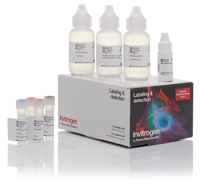
Alexa Fluor™ 594 Tyramide SuperBoost™ Kit, goat anti-rabbit IgG
SuperBoost™チラミドシグナル増幅は、多重化可能な蛍光免疫細胞化学(ICC)、免疫組織化学(IHC)、およびin situハイブリダイゼーション(ISH詳細を見る


製品番号(カタログ番号) B40925
価格(JPY)お問い合わせください ›
162,500
Each
数量:
150枚のスライドに十分な量
製品タイプ:
Tyramide Kit
SuperBoost™チラミドシグナル増幅は、多重化可能な蛍光免疫細胞化学(ICC)、免疫組織化学(IHC)、およびin situハイブリダイゼーション(ISH)において、少量のターゲットを検出するためのもっとも感度の高い方法です。SuperBoostキットは、AlexaFluor™色素の輝度と、ポリ-HRPを介したチラミド標識反応の優れたシグナル増幅を組み合わせることで、標準的な手法に比べ、10~200倍高い感度を実現します。SuperBoostキットの感度は、さらに、TSA™のような通常のチラミド増幅技術の2-10倍高くなっています。優れた研究のために、SuperBoostキットは結果をシャープにし、標準的なイメージング法では明らかにできない重要な領域を明確に可視化します。
SuperBoostキットは使いやすく、あらゆる細胞または組織タイプを使用して、標準的なICC、IHC、またはFISH実験プロトコルに簡単に適応できます。SuperBoostキットを使用してラベリングされた細胞は、あらゆるタイプの顕微鏡を使用してイメージングでき、高解像度のマルチプレックス画像を生成することができます。この特別なキットは、標準のRed/Texas Red™フィルターキューブを使用して検出されたAlexaFluor 594チラミド(591/617 ex/em)を特徴としています。このキットには、ポリHRP標識ヤギ抗ウサギIgG二次抗体も含まれています。
SuperBoostキットの特長は以下のとおりです。
•蛍光イメージングによる低濃度または検出困難なターゲットの検出に優れた感度
•シンプルなプロトコルと標準フィルターを使用した検出
•高解像度のマルチプレックス画像に適しています—DAPI、二次抗体、およびその他のSuperBoostキットとの共標識
•標準的なICC/IHC/ISH実験の10-100分の1の一次抗体が必要です
SuperBoostキットは、西洋ワサビペルオキシダーゼ(HRP)の触媒活性を利用して、標的タンパク質または核酸配列の高密度標識をin situで生成する、チラミドシグナル増幅システムに基づいています。SuperBoostキットを使用した一般的なICC/IHC/ISH実験では、標準的なICC/IHC/ISH実験に比べ、必要な一次抗体が10-100倍少なくて済みます。SuperBoostキットは、特定の標識の濃度をバックグラウンドに対して大きく高めるため、高い内因性自家蛍光が検出されるサンプルで特定の標識を検出するように、プロトコルを容易に最適化できます。
SuperBoostキットの利点
Alexa Fluorチラミドを使用した標識の機能向上には、次のようなものがあります。SuperBoostキットはAlexa Fluorチラミドを使用します。これはHRPと反応して、周囲のタンパク質や他の類似分子に明るい光安定性のAlexa Fluor色素を究極的に沈着させます。SuperBoostキットは、Alexa Fluor色素の輝度と、チラミドシグナル増幅の強化を組み合わせて優れたシグナルを生成する唯一のキットです。
ポリ-HRPの強化:TSAとは異なり、SuperBoostキットはポリ-HRP標識二次抗体を使用します。このようなシステムでは、いくつかのHRP酵素が短いポリマーと接合し、通常のHRPシステムよりも数倍多くシグナルを増強します。ポリHRPは、抗体が通常のHRP標識二次抗体と同様に効率的に細胞または組織を貫通するように構造化されています。モル酵素/抗体タンパク質比の平均値は「4」です。
反応停止液:他の酵素ベースのラベリングシステムと同様に、シグナルを過剰に発生させる可能性があります。SuperBoostキットには、HRP反応を停止させるHRP停止液が含まれています。HRP停止液を使用すると、バックグラウンド標識を増加させることなく、最大の標識を得ることができます。最適化されたHRP反応時間で生成されたイメージと、標準的なICC/IHC/ISH手法で生成されたイメージとでは、鮮明さは同程度ですが、感度は前者のほうが10-200倍高くなります。
バックグラウンドの低減:SuperBoostキットには、内因性ペルオキシダーゼおよび蛍光バックグラウンド標識を除去または低減するためのブロッカーが含まれています。これらのブロッカーは、特定の標識のみを増幅し、その他の標識やバックグラウンド標識は低量に抑えます。
研究用途にのみご使用ください。診断目的には使用できません。
仕様
製品タイプTyramide Kit
数量150枚のスライドに十分な量
品質保持期間6カ月
出荷条件室温またはウェットアイスでの出荷
コンジュゲートAlexa Fluor 594
製品ラインSuperBoost™
Unit SizeEach
組成および保存条件
顕微鏡スライド(18 x 18 mm)150枚に十分なキット1個、以下が含まれます:ブロッキングバッファー(1X)、22.5 mLポリHRP標識ヤギ抗ウサギ二次抗体(1X)、22.5 mL Alexa Fluorチラミド試薬 過酸化水素