Search Thermo Fisher Scientific

DualBeam Microscopes
Helios 5 Hydra DualBeam
PFIB SEM with multiple ion species for 3D EM and TEM sample preparation.
Join the Conversation
cd49 - Key/Resources/Appl/Tech/Doc/Contact
PFIB SEM with the Helios 5 Hydra DualBeam
The Thermo Scientific Helios 5 Hydra DualBeam (plasma focused ion beam scanning electron microscope, PFIB-SEM) is a versatile multi-application tool that has four different ion species (argon, nitrogen, oxygen, and xenon), allowing you to choose the ions that provide the best results for samples including metals,batteries, semiconductors, fiber composites, and biological tissues. Great results start with sample preparation whether you are performing scanning transmission electron microscopy (STEM) and transmission electron microscopy (TEM) sample preparation, 3D characterization, large area failure analysis, volume electron microscopy or cellular cryo-tomography.
You can switch easily between argon, nitrogen, oxygen, and xenon in under ten minutes without sacrificing performance. This unprecedented flexibility significantly expands the potential application space of PFIB and enables research of ion-sample interactions to optimize existing use cases.
The Helios 5 Hydra DualBeam combines the new, innovative multi-ion-species plasma FIB (PFIB) column with the monochromated Thermo Scientific Elstar UC+ SEM Column to provide advanced focused ion- and electron-beam performance. Intuitive software and an unprecedented level of automation and ease-of-use allow observation and analysis of relevant subsurface volumes.
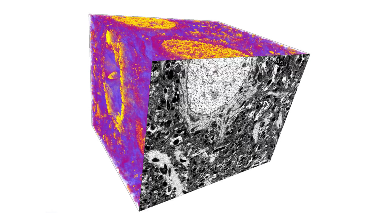
3D reconstruction of mouse brain tissue embedded in LR White (acrylic-based) resin acquired with an O+ ion beam and Auto Slice and View Software.
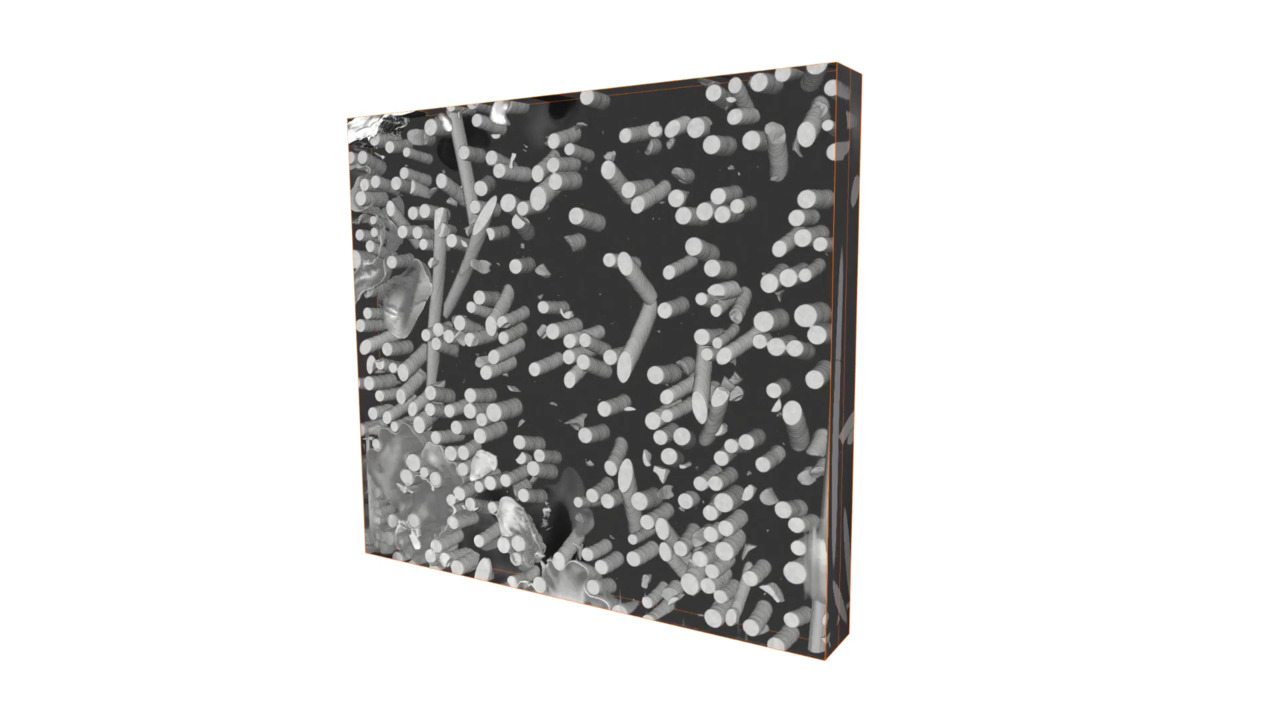
3D reconstruction of an automotive oil filter casing (polymer/glass fiber composite) acquired with an O+ ion beam and Auto Slice and View Software.
3D Reconstruction of High Bandwidth Memory Package by Plasma FIB Slice and View

Multiple plasma ion species
Allows multi-application labs to quickly switch between four ion species (Xe, Ar, O, and N) to optimize for samples ranging from silicon and metals to cells

Ga+ Free TEM
Facilitates fast and easy preparation of high-quality samples, including Ga+ free TEM, atom probe tomography, and resin-embedded samples

3D tomography
Performs high-resolution, large volume, 3D tomography and combines multi-modal subsurface 3D information (SEM, EDS, EBSD)

Cryogenic conditions
Delivers high-resolution SEM images with high contrast even at cryogenic conditions for biological samples

Precise milling control
Provides precise control of your milling experiments with time-saving automated software and workflow-based solutions
Multiple ion species for optimized PFIB results
The Helios 5 Hydra DualBeam is an all-in-one instrument with a unique multiple ion species PFIB column with four ion species (Xe, Ar, O, N) to be used as a primary beam with a patented, automated, fast, and easy switching capability (<10 minutes). The Helios 5 Hydra PFIB is Ga-ion free, which is vital for sensitive samples such as aluminum-containing materials or compound semiconductors, like Sic or GaN. The multiple ion options increase throughout and do not require complex accessories to improve cut face quality (e.g., milling curtain mitigation) for a wide range of materials.

Click image to enlarge

Click image to enlarge

Click image to enlarge
Lamellae preparation with AutoTEM Software
High-quality sample preparation is critical for successful high-resolution analysis with STEM or TEM. Conventional methods used to prepare ultra-thin samples required for S/TEM are slow and can require many hours, or even days, of effort by highly trained personnel. This is further complicated by the variety of materials and the need for site-specific information. Plasma FIB, coupled with optional AutoTEM Software, resolves many of these challenges.
Serial section electron microscopy with Auto Slice & View Software
Combined with optional Auto Slice & View Software, the Helios Hydra DualBeam offers high throughput and fully automated 3D data collection, even at cryogenic temperatures. Large volume analysis can be limited by cross-sectional information alone, but 3D characterization of buried features for failure analysis or metrology provides a complete picture. The automated workflow enhances throughput and ease of use, even for challenging materials with complex structures or defects.


Click image to enlarge
PFIB spin mill for large planar milling and imaging
The optional Spin Mill method on the Helios 5 Hydra DualBeam provides large-area planar milling up to 1 mm in diameter for polished surface imaging, 3D characterization, or buried defect inspection. The Spin Mill process is fully automated and easy to set up using Auto Slice & View Software. A key advantage is the range of materials where this method is effective. Spin mill can be a preparation method for advanced semiconductor device delayering and inspection workflows, allowing removal of thick metallization. For resin-embedded life science samples, the spin mill method can be used to ablate the sample over a large area to locate and image target regions at high resolution

Click image to enlarge

Click image to enlarge

Click image to enlarge
PFIB Delayering for inspection and failure analysis
The unique combination of Helios 5 Hydra xenon plasma FIB technology and proprietary Thermo Scientific Dx chemistry enables damage-free delayering of semiconductor devices, including advanced logic samples and 3D NAND memory devices. Automatic de-processing allows access to buried information for advanced devices that would otherwise be unattainable. Thermo Fisher Scientific's unique Plasma FIB delayering process complements the nProber IV and Hyperion II, delivering a robust nanoprobing and transistor characterization workflow.

Click image to enlarge
Fast milling for large volume multi-modal 3D tomography
Subsurface or three-dimensional characterization is often required to better understand material properties. In many cases, large volumes inaccessible by conventional Ga+ FIB instruments are necessary to obtain representative and relevant results. The excellent high-current performance of the Helios 5 Hydra PFIB with optional Thermo Scientific Auto Slice & View Software empowers the fully automated acquisition of large volume 3D datasets in a multitude of modalities. This includes backscattered electron (BSE) imaging for maximum materials contrast, energy dispersive spectroscopy (EDS) for compositional information, and electron backscatter diffraction (EBSD) for crystallographic information.

Click image to enlarge
3D imaging at cryogenic temperatures
The Helios 5 Hydra DualBeam can be equipped with an optional cryo-stage that can be used for dedicated cryo-lamellae preparation with AutoTEM Cryo or cryo-volume imaging with Auto Slice & View Software. With the Elstar SEM Column, excellent contrast is achievable to uncover sub-cellular details on high pressure frozen and plunge frozen samples. No staining is required.
Learn more about the capabilities of the Hydra Bio Plasma-FIB ›
")
Click image to enlarge
Fluorescence light microscope correlative system (iFLM)
The optional iFLM Correlative System is an integrated light microscope for cryo-correlative imaging inside the Helios 5 Hydra PFIB high-vacuum chamber allowing you to combine fluorescence imaging and ion milling within a single cryo-DualBeam microscope. Read the iFLM Correlative System datasheet.
Datasheets


Applications notes


Infographics


Related FIB SEM DualBeam systems and resources


Webinars
Style Sheet for Products Table Specifications
Style Sheet for Komodo Tabs
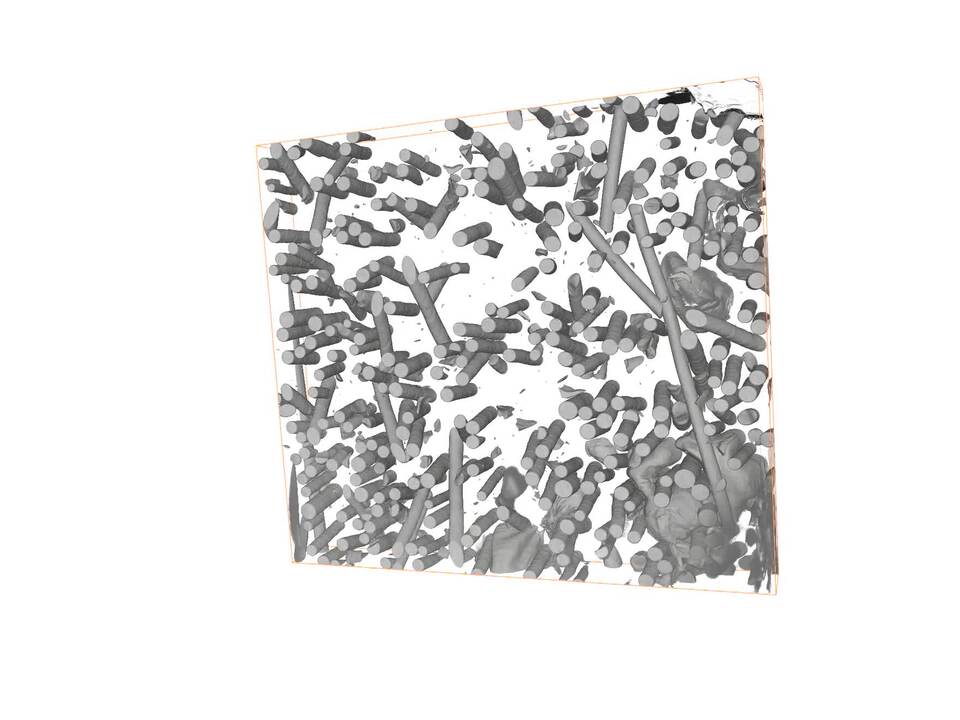
3D reconstruction of an automotive oil filter casing, acquired with the Helios Hydra DualBeam and Auto Slice & View 4 Software for automated serial sectioning. Horizontal field width = 350 µm.
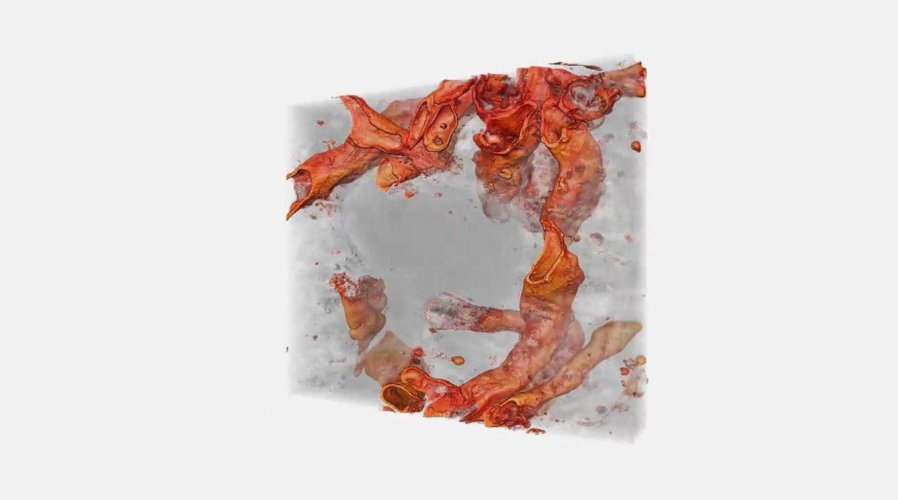
3D reconstruction of a mouse brain tissue acquired with a Helios Hydra UX DualBeam using O+ focused ion beam. Sample preparation: Conventional cemical fixation. Epon resin. 721 slices (thickness ~ 4 nm). HFW 16.4 µm (xy resolution 5x4 nm). Acquisition time ~ 17 h.

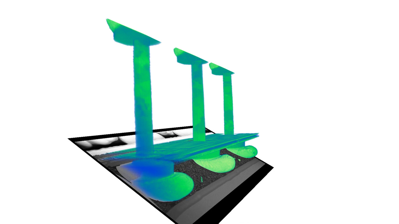
Hydra's unique multi-ion beam technology enables the precise and efficient analysis of buried defects in semiconductor packaged die and power devices
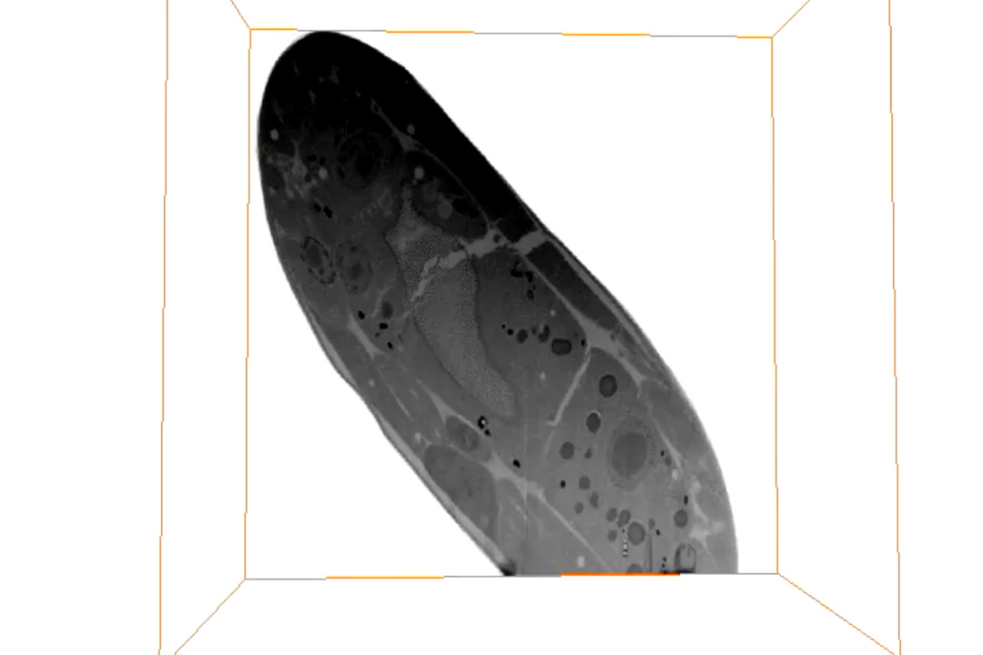
3D reconstruction of Caenorhabditis elegans L1 with a Helios Hydra CX DualBeam using O+ focused ion beam. Sample preparation: high pressure frozen and freeze substituted and HM20 embedded. Tomogram of a 30 × 30 × 15 µm (resolution ~10 x ×10 x ×15 nm). Acquisition time ~ 16 h. Courtesy Dr. Alex DeMarco, Monash University, Australia.
https://www.biorxiv.org/content/10.1101/457820v1.full.pdf
Publications
3D reconstruction of an automotive oil filter casing, acquired with the Helios Hydra DualBeam and Auto Slice & View 4 Software for automated serial sectioning. Horizontal field width = 350 µm.
3D reconstruction of a mouse brain tissue acquired with a Helios Hydra UX DualBeam using O+ focused ion beam. Sample preparation: Conventional cemical fixation. Epon resin. 721 slices (thickness ~ 4 nm). HFW 16.4 µm (xy resolution 5x4 nm). Acquisition time ~ 17 h.
Hydra's unique multi-ion beam technology enables the precise and efficient analysis of buried defects in semiconductor packaged die and power devices
3D reconstruction of Caenorhabditis elegans L1 with a Helios Hydra CX DualBeam using O+ focused ion beam. Sample preparation: high pressure frozen and freeze substituted and HM20 embedded. Tomogram of a 30 × 30 × 15 µm (resolution ~10 x ×10 x ×15 nm). Acquisition time ~ 16 h. Courtesy Dr. Alex DeMarco, Monash University, Australia.
https://www.biorxiv.org/content/10.1101/457820v1.full.pdf
Publications

Process control using electron microscopy
Modern industry demands high throughput with superior quality, a balance that is maintained through robust process control. SEM and TEM tools with dedicated automation software provide rapid, multi-scale information for process monitoring and improvement.

Quality control and failure analysis
Quality control and assurance are essential in modern industry. We offer a range of EM and spectroscopy tools for multi-scale and multi-modal analysis of defects, allowing you to make reliable and informed decisions for process control and improvement.

Fundamental Materials Research
Novel materials are investigated at increasingly smaller scales for maximum control of their physical and chemical properties. Electron microscopy provides researchers with key insight into a wide variety of material characteristics at the micro- to nano-scale.

Semiconductor Failure Analysis
Complex semiconductor device structures result in more places for defects to hide. Learn more about failure analysis solutions to isolate, analyze, and repair defects.

Cryo-Tomography
Cryo-electron tomography (cryo-ET) delivers both structural information about individual proteins as well as their spatial arrangements within the cell. This makes it a truly unique technique and also explains why the method has such an enormous potential for cell biology. Cryo-ET can bridge the gap between light microscopy and near-atomic-resolution techniques like single-particle analysis.

Volume Electron Microscopy
The emerging field of volume EM refers to a variety of imaging approaches and processing techniques that uses electron microscopy to explore below the surface of cellular ultrastructure, tissue, and small model organisms in 3D, at micron to millimeter volume scales, at nanometer-level resolutions, and even at native state under cryogenic conditions.

3D Materials Characterization
Development of materials often requires multi-scale 3D characterization. DualBeam instruments enable serial sectioning of large volumes and subsequent SEM imaging at nanometer scale, which can be processed into high-quality 3D reconstructions of the sample.

(S)TEM Sample Preparation
DualBeam microscopes enable the preparation of high-quality, ultra-thin samples for (S)TEM analysis. Thanks to advanced automation, users with any experience level can obtain expert-level results for a wide range of materials.

APT Sample Preparation
Atom probe tomography (APT) provides atomic-resolution 3D compositional analysis of materials. Focused ion beam (FIB) microscopy is an essential technique for high-quality, orientation, and site-specific sample preparation for APT characterization.

Cross-sectioning
Cross sectioning provides extra insight by revealing sub-surface information. DualBeam instruments feature superior focused ion beam columns for high-quality cross sectioning. With automation, unattended high-throughput processing of samples is possible.

In Situ experimentation
Direct, real-time observation of microstructural changes with electron microscopy is necessary to understand the underlying principles of dynamic processes such as recrystallization, grain growth, and phase transformation during heating, cooling, and wetting.
Multi-scale analysis
Novel materials must be analyzed at ever higher resolution while retaining the larger context of the sample. Multi-scale analysis allows for the correlation of various imaging tools and modalities such as X-ray microCT, DualBeam, Laser PFIB, SEM and TEM.

Device Delayering
Shrinking feature size, along with advanced design and architecture, results in increasingly challenging failure analysis for semiconductors. Damage-free delayering of devices is a critical technique for the detection of buried electrical faults and failures.
Cryo-Tomography
Cryo-electron tomography (cryo-ET) delivers both structural information about individual proteins as well as their spatial arrangements within the cell. This makes it a truly unique technique and also explains why the method has such an enormous potential for cell biology. Cryo-ET can bridge the gap between light microscopy and near-atomic-resolution techniques like single-particle analysis.
Volume Electron Microscopy
The emerging field of volume EM refers to a variety of imaging approaches and processing techniques that uses electron microscopy to explore below the surface of cellular ultrastructure, tissue, and small model organisms in 3D, at micron to millimeter volume scales, at nanometer-level resolutions, and even at native state under cryogenic conditions.
3D Materials Characterization
Development of materials often requires multi-scale 3D characterization. DualBeam instruments enable serial sectioning of large volumes and subsequent SEM imaging at nanometer scale, which can be processed into high-quality 3D reconstructions of the sample.
(S)TEM Sample Preparation
DualBeam microscopes enable the preparation of high-quality, ultra-thin samples for (S)TEM analysis. Thanks to advanced automation, users with any experience level can obtain expert-level results for a wide range of materials.
APT Sample Preparation
Atom probe tomography (APT) provides atomic-resolution 3D compositional analysis of materials. Focused ion beam (FIB) microscopy is an essential technique for high-quality, orientation, and site-specific sample preparation for APT characterization.
Cross-sectioning
Cross sectioning provides extra insight by revealing sub-surface information. DualBeam instruments feature superior focused ion beam columns for high-quality cross sectioning. With automation, unattended high-throughput processing of samples is possible.
In Situ experimentation
Direct, real-time observation of microstructural changes with electron microscopy is necessary to understand the underlying principles of dynamic processes such as recrystallization, grain growth, and phase transformation during heating, cooling, and wetting.
Multi-scale analysis
Novel materials must be analyzed at ever higher resolution while retaining the larger context of the sample. Multi-scale analysis allows for the correlation of various imaging tools and modalities such as X-ray microCT, DualBeam, Laser PFIB, SEM and TEM.
Device Delayering
Shrinking feature size, along with advanced design and architecture, results in increasingly challenging failure analysis for semiconductors. Damage-free delayering of devices is a critical technique for the detection of buried electrical faults and failures.
Style Sheet to change H2 style to p with em-h2-header class
Electron microscopy services for
the materials science
To ensure optimal system performance, we provide you access to a world-class network of field service experts, technical support, and certified spare parts.
Electron microscopy support and resources
Style Sheet for Support and Service footer
Style Sheet for Fonts
Style Sheet for Cards