Search Thermo Fisher Scientific

Materials Science
3D Electron Microscopy
3D electron microscopy through focused ion beam milling and scanning electron microscopy imaging.
Join the conversation
48ab - Appl/Samples/Products/Resources/Contact
3D electron microscopy
Whether studying new materials or attempting to find the root causes of failures, highly localized characterization of increasingly complex samples with ever-smaller features is becoming increasingly critical. In fact, sub-surface or three-dimensional characterization is often required to fully understand the material properties of a sample. This can be particularly useful for heterogeneous materials or the detection and characterization of inclusions and precipitates. DualBeam (focused ion beam – scanning electron microscopy, FIB-SEM) instrumentation enables the acquisition of high-quality 3D datasets, at nanometer resolution, by serial sectioning (sequential application of SEM imaging and FIB milling).
Plasma FIB (PFIB)
Often, large volumes, which are inaccessible with conventional gallium FIB instruments, are necessary to obtain representative and statistically relevant results due to the additional contextual information a large sample provides. Plasma FIB instruments, such as the Thermo Scientific Helios 5 PFIB DualBeam and the Thermo Scientific Helios Hydra DualBeam, offer excellent high-current performance enabling fully automated, high-quality, large-volume 3D datasets in a variety of modalities. Additionally, the multiple ion source technology of the Helios Hydra DualBeam allows you to choose the best ion source for each particular sample and use case.
Plasma FIB serial sectioning can provide information on materials contrast (with backscattered electron imaging), compositional information (with energy dispersive spectroscopy), and microstructural and crystallographic information (with electron backscatter diffraction). This data can then be visualized with Thermo Scientific Avizo Software, delivering a unique workflow solution for the highest-resolution, advanced 3D characterization and analysis at the nanometer scale.
Laser PFIB
The Thermo Scientific Helios 5 Laser PFIB System goes beyond regular plasma FIB to perform cross-sectioning and 3D characterization of even larger volumes, up to millimeter-scale (with >15,000x faster coarse material removal as compared to gallium FIB). It also facilitates the processing of materials that are typically challenging for ion beam milling, such as charging and beam sensitive materials, with minimal damage.

3D EBSD reconstruction of a zircalloy sample (250 x 250 x 220 μm³) produced with the Helios PFIB DualBeam, Auto Slice & View 4 Software, and Avizo Software.

Large volume femto-second laser tomography of a carbon-fiber reinforced composite acquired with Laser PFIB on the Helios DualBeam. Horizontal field width is 500 µm.

Large-area cross-section of a steel sample manufactured by selective laser melting technology. Total milling time by laser is <5 minutes (area: 600 x 350 µm). Femto-second laser processing results in high-quality cut face, which directly allows EBSD mapping without the need for post-cleaning with FIB.

Automotive paint scratch testing. High throughput, large-scale cross-sectional imaging was performed with the Helios Plasma FIB DualBeam.

3D reconstruction of W-Mo-Cu sample using a combination of BSE (green-blue) and EDS (orange) data, which has been produced with a Thermo Scientific Scios 2 DualBeam System, Auto Slice and View 4 and Avizo for Materials Science software.
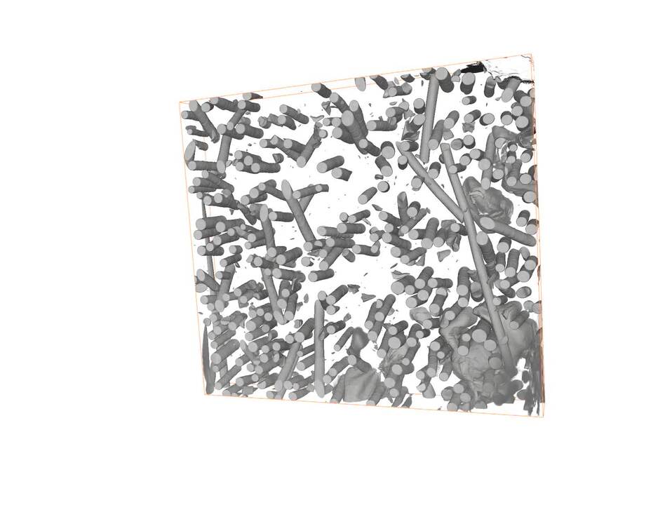
Automotive oil filter casing (a polymer/glass fiber composite) imaged on the Helios Hydra DualBeam and reconstructed into a 3D volume in Avizo Software. Horizontal field width is 350 µm.
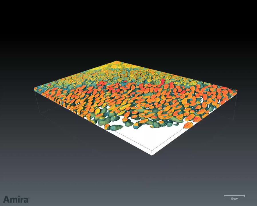
Embedded tissue sample imaged on the Helios Hydra DualBeam and reconstructed into a 3D volume in Amira Software, highlighting the ability of the Helios Hydra DualBeam to perform high-quality 3D characterization of organic materials.
Large volume femto-second laser tomography of a carbon-fiber reinforced composite acquired with Laser PFIB on the Helios DualBeam. Horizontal field width is 500 µm.
Large-area cross-section of a steel sample manufactured by selective laser melting technology. Total milling time by laser is <5 minutes (area: 600 x 350 µm). Femto-second laser processing results in high-quality cut face, which directly allows EBSD mapping without the need for post-cleaning with FIB.
Automotive paint scratch testing. High throughput, large-scale cross-sectional imaging was performed with the Helios Plasma FIB DualBeam.
3D reconstruction of W-Mo-Cu sample using a combination of BSE (green-blue) and EDS (orange) data, which has been produced with a Thermo Scientific Scios 2 DualBeam System, Auto Slice and View 4 and Avizo for Materials Science software.
Automotive oil filter casing (a polymer/glass fiber composite) imaged on the Helios Hydra DualBeam and reconstructed into a 3D volume in Avizo Software. Horizontal field width is 350 µm.
Embedded tissue sample imaged on the Helios Hydra DualBeam and reconstructed into a 3D volume in Amira Software, highlighting the ability of the Helios Hydra DualBeam to perform high-quality 3D characterization of organic materials.

Quality control and failure analysis
Quality control and assurance are essential in modern industry. We offer a range of EM and spectroscopy tools for multi-scale and multi-modal analysis of defects, allowing you to make reliable and informed decisions for process control and improvement.

Fundamental Materials Research
Novel materials are investigated at increasingly smaller scales for maximum control of their physical and chemical properties. Electron microscopy provides researchers with key insight into a wide variety of material characteristics at the micro- to nano-scale.

Battery Research
Battery development is enabled by multi-scale analysis with microCT, SEM and TEM, Raman spectroscopy, XPS, and digital 3D visualization and analysis. Learn how this approach provides the structural and chemical information needed to build better batteries.

Polymers Research
Polymer microstructure dictates the material’s bulk characteristics and performance. Electron microscopy enables comprehensive microscale analysis of polymer morphology and composition for R&D and quality control applications.

Metals Research
Effective production of metals requires precise control of inclusions and precipitates. Our automated tools can perform a variety of tasks critical for metal analysis including; nanoparticle counting, EDS chemical analysis and TEM sample preparation.

Oil and Gas
As the demand for oil and gas continues, there is an ongoing need for efficient and effective extraction of hydrocarbons. Thermo Fisher Scientific offers a range of microscopy and spectroscopy solutions for a variety of petroleum science applications.

Fibers and Filters
The diameter, morphology and density of synthetic fibers are key parameters that determine the lifetime and functionality of a filter. Scanning electron microscopy (SEM) is the ideal technique for quickly and easily investigating these features.

Geological Research
Geoscience relies on consistent and accurate multi-scale observation of features within rock samples. SEM-EDS, combined with automation software, enables direct, large-scale analysis of texture and mineral composition for petrology and mineralogy research.
Style Sheet for Instrument Cards Original


Style Sheet for Komodo Tabs
Style Sheet to change H2 style to p with em-h2-header class
Style Sheet for Support and Service footer
Style Sheet for Fonts
Style Sheet for Cards
Electron microscopy services for
the materials science
To ensure optimal system performance, we provide you access to a world-class network of field service experts, technical support, and certified spare parts.