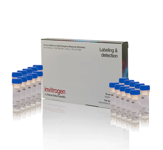
CoroNa™ Green, AM, cell permeant - Special Packaging
Invitrogen™

CoroNa™ Green, AM, cell permeant - Special Packaging

CoroNa Green dye is a sodium ion indicator that exhibits an increase in green fluorescence emission intensity upon binding Na+,Read more
Promo ImagePromotion
Promo code:P5906365
Don't miss out: buy 3, only pay for 2
Promo ImagePromotion
Promo code:RPUZZ25
Stock up on essentials to piece your discovery together
Have Questions?
Catalog NumberQuantity
C36676Promo Image20 x 50 μg
Catalog number C36676
Price (USD)
775.65
Online Exclusive
844.00
Save 68.35 (8%)
Each
In stock
Add to cart
Quantity:
20 x 50 μg
Recurring order eligible. Learn more »
Price (USD)
775.65
Online Exclusive
844.00
Save 68.35 (8%)
Each
Add to cart
Ask our AI about this Product
CoroNa Green dye is a sodium ion indicator that exhibits an increase in green fluorescence emission intensity upon binding Na+, with little shift in wavelength. Like our other sodium indicators, SBFI (Cat. nos. S1262, S1263, S1264) and Sodium Green dye (Cat. nos. S6900, S6901), the CoroNa Green indicator allows spatial and temporal resolution of Na+ concentrations in the presence of physiological concentrations of other monovalent cations. CoroNa Green AM dye loads into cells more efficiently than does Sodium Green tetraacetate, and the CoroNa Green indicator responds to a broader range of Na+ concentration. With absorption/emission maxima of ∼492/516 nm for the Na+-bound form, CoroNa Green dye can be detected by any instrument that can detect fluorescein. CoroNa Green dye is also available in a cell impermeant form (Cat. no. C36675).

Learn more about ion indicators including calcium, potassium, pH, and membrane potential indicators ›

For Research Use Only. Not for use in diagnostic procedures.
Specifications
Detection MethodFluorescence
Dye TypeSodium Indicator
Quantity20 x 50 μg
Shipping ConditionRoom Temperature
For Use With (Application)Cell Viability and Proliferation
For Use With (Equipment)Fluorescence Microscope
Product LineCoroNa
Product TypeSodium Ion Indicator
Unit SizeEach
Contents & Storage
Store in freezer -5°C to -30°C and protect from light.
Have questions about this product? Ask our AI assisted search.
Why don't I see a significant change in signal for my live-cell fluorescent indicator dye?
This is an AI-powered search and may not always get things right. You can help us make it better with a thumbs up or down on individual answers or by selecting the “Give feedback" button. Your search history and customer login information may be retained by Thermo Fisher and processed in accordance with our Privacy Notice.

Customers who viewed this item also viewed


Documents & Downloads

Certificates

Lot #Certificate TypeDateCatalog Number(s)
2935414Certificate of AnalysisAug 23, 2024C36676
2892724Certificate of AnalysisMar 01, 2024C36676
2738346Certificate of AnalysisSep 29, 2023C36676
2513382Certificate of AnalysisDec 30, 2022C36676
2339882Certificate of AnalysisJul 03, 2021C36676
5 results displayed, search above for a specific certificate

Safety Data Sheets

Frequently asked questions (FAQs)

Regardless of the type of live-cell indicator dye (e.g., calcium indicators, pH indicator, metal ion indicators), make sure there is no serum during the loading step, which can prematurely cleave dyes with AM esters and bind dyes non-specifically. Always optimize the dye concentration and staining time with a positive control before you run your test samples, to give the best signal-to-background. Always run a positive control with a buffer containing free ions of known concentration and an ionophore to open pores to those ions (for instance, for calcium indicators like Fluo-4 AM, this would include a buffer with added calcium combined with calcimycin, or for pH indicators, buffers of different pHs combined with nigericin). Reactive oxygen indicators, such as CellROX Green or H2DCFDA would require a cellular reactive oxygen species (ROS) stimulant as a positive control, such as menadione. Finally, make sure your imaging system has a sensitive detector. Plate readers, for instance, have much lower detector efficiency over background, compared to microscopy or flow cytometry.

Find additional tips, troubleshooting help, and resources within our Cell Analysis Support Center.

Citations & References (25)

Citations & References
Abstract
Arabidopsis fatty acid desaturase FAD2 is required for salt tolerance during seed germination and early seedling growth.
Authors:Zhang J, Liu H, Sun J, Li B, Zhu Q, Chen S, Zhang H,
Journal:PLoS One
PubMed ID:22279586
'Fatty acid desaturases play important role in plant responses to abiotic stresses. However, their exact function in plant resistance to salt stress is unknown. In this work, we provide the evidence that FAD2, an endoplasmic reticulum localized ?-6 desaturase, is required for salt tolerance in Arabidopsis. Using vacuolar and plasma ... More
Diacylglycerol analogues activate second messenger-operated calcium channels exhibiting TRPC-like properties in cortical neurons.
Authors:Tu P, Kunert-Keil C, Lucke S, Brinkmeier H, Bouron A,
Journal:J Neurochem
PubMed ID:19094061
'The lipid diacylglycerol (DAG) analogue 1-oleoyl-2-acetyl-sn-glycerol (OAG) was used to verify the existence of DAG-sensitive channels in cortical neurons dissociated from E13 mouse embryos. Calcium imaging experiments showed that OAG increased the cytosolic concentration of Ca(2+) ([Ca(2+)]i) in nearly 35% of the KCl-responsive cells. These Ca(2+) responses disappeared in a ... More
Membrane phosphatidylserine distribution as a non-apoptotic signalling mechanism in lymphocytes.
Authors:Elliott JI, Surprenant A, Marelli-Berg FM, Cooper JC, Cassady-Cain RL, Wooding C, Linton K, Alexander DR, Higgins CF
Journal:Nat Cell Biol
PubMed ID:16025105
'Phosphatidylserine (PS) exposure is normally associated with apoptosis and the removal of dying cells. We observed that PS is exposed constitutively at high levels on T lymphocytes that express low levels of the transmembrane tyrosine phosphatase CD45RB. CD45 was shown to be a negative regulator of PS translocation in response ... More
Loss of halophytism by interference with SOS1 expression.
Authors:Oh DH, Leidi E, Zhang Q, Hwang SM, Li Y, Quintero FJ, Jiang X, D'Urzo MP, Lee SY, Zhao Y, Bahk JD, Bressan RA, Yun DJ, Pardo JM, Bohnert HJ,
Journal:Plant Physiol
PubMed ID:19571313
'The contribution of SOS1 (for Salt Overly Sensitive 1), encoding a sodium/proton antiporter, to plant salinity tolerance was analyzed in wild-type and RNA interference (RNAi) lines of the halophytic Arabidopsis (Arabidopsis thaliana)-relative Thellungiella salsuginea. Under all conditions, SOS1 mRNA abundance was higher in Thellungiella than in Arabidopsis. Ectopic expression of ... More
Cationic gradient reversal and cytoskeleton-independent volume regulatory pathways define an early stage of apoptosis.
Authors:Bortner CD, Sifre MI, Cidlowski JA,
Journal:J Biol Chem
PubMed ID:18187415
'Cell shrinkage, or apoptotic volume decrease (AVD), is a ubiquitous characteristic of programmed cell death that is independent of the death stimulus and occurs in all examples of apoptosis. Here we distinguished two specific stages of AVD based on cell size and a unique early reversal of intracellular ions that ... More
25 total citations

Other products to consider


Share catalog number, name or link

1x1 image pixel for data collection