Search Thermo Fisher Scientific
- Contáctenos
- Orden Rápida
-
¿No tiene una cuenta? Crear una cuenta
Search Thermo Fisher Scientific

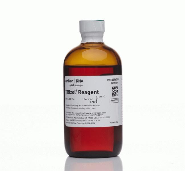


| Número de catálogo | Cantidad |
|---|---|
| 15596018 | 200 mL |
| 15596026 | 100 mL |
Even the most experienced molecular biologist has screamed their favorite expletive during an RNA extraction. To a non-scientist, it can look like this poor person is losing it, yelling at an innocent tube, filled with clear, nondescript liquid. Read more ›
SDSDegraded RNA can cause an increased absorbance at 260 nm.
Find additional tips, troubleshooting help, and resources within ourRNA Sample Collection, Protection, and Isolation Support Center.
This is most likely polysaccharides or cell membranes; DNA should be in the interphase. In samples containing blood (e.g., liver), a red viscous layer may be visible on top of the pellet. This is most likely due to blood products and should not be carried over with the supernatant.
- This is common with skin samples. It is assumed that there is fat in these samples, and the fat micelles float during the centrifugation. In skin samples, the micelles pick up melanin pigment and cause the aqueous phase to appear colored. Fat micelles may also pick up pigment from the TRIzol Reagent itself and cause a pinkish color. If a sample is thought to contain fat, the sample homogenate in TRIzol Reagent may be centrifuged prior to addition of chloroform. The fat will appear as a clear layer at the top of the supernatant; this should be pipetted off and discarded.
- If a sample contains a lot of blood, the aqueous phase may appear cloudy and/or yellowish (this may be due to iron in the hemoglobin). If the centrifuge used is not cold, the organic phase will be a deeper maroon color; some of this color may come into the aqueous phase and cause it to appear orange or yellow.
- A pinkish aqueous phase may also be caused by overdilution of the sample (i.e., a sample to TRIzol Reagent ratio > 1:10), as well as too much salt or protein in the sample. This can cause premature phase separation, which can be remedied by adding a bit more TRIzol Reagent to the sample. If the RNA is isolated from a pinkish aqueous phase, chances are that it will be contaminated with DNA.
These are our recommendations:
1. Upstream tissue procurement and tissue specimen preparation—if possible, fix tissues within one hour of surgical resection. Extensive degradation of RNA can occur before completion of the fixation process. The optimal fixation time is 12-24 hours, using neutral-buffered formalin or paraformaldehyde. Fixed tissues should be thoroughly dehydrated prior to the embedding process.
2. Block storage—storage of blocks without cut faces, when possible, prevents ongoing damage from exposure to atmospheric oxygen, water, and other environmental factors such as light and infestation (fungus, insects, etc.).
3. Choice of tissue type, size, and amount being used for RNA isolation—the recommended tissue thickness is 10-20 µm. The number of sections used is determined by the tissue type (which impacts cell density) and surface area (recommended size: 50-300 mm2). Excess starting material can cause filter clogging, resulting in poor yield.
4. Avoid using an excessive amount of paraffin for embedding tissues—when possible, excess paraffin should be trimmed away prior to starting the purification protocol. For xylene-based purification methods, two xylene treatments at room temperature should be sufficient for complete deparaffinization. If desired, you can perform a more rigorous 37-55 degrees C treatment for up to 30 minutes. After the xylene deparaffinization, it is crucial that the 100% ethanol is completely removed and the pellets are dry after the two 100% ethanol washes. The magnetic bead method employs novel chemistries to deal with the paraffin that limits input to 20 µm sections.
Read more about RNA isolation from FFPE tissues here (https://www.thermofisher.com/us/en/home/life-science/dna-rna-purification-analysis/rna-extraction/rna-sample-extraction/working-with-ffpe-samples.html).
If a sample is known to have a high content of proteoglycans and/or polysaccharides (such as rat liver, rat aorta, plants), the following modification of the RNA precipitation step should remove these contaminating compounds from the isolated RNA:
- Add 0.25 mL of isopropanol to the aqueous phase followed by 0.25 mL of a high-salt precipitation solution (0.8 M sodium citrate and 1.2 M NaCl; no pH adjustment necessary) per 1 mL of TRIzol Reagent used for homogenization. Mix the resulting solution, centrifuge, and proceed with isolation as described in the protocol.
This modified precipitation effectively precipitates RNA and maintains proteoglycans and polysaccharides in a soluble form. To isolate pure RNA from plant material containing a very high level of polysaccharides, the modified precipitation should be combined with an additional centrifugation of the initial homogenate. In general, we do not recommend high-salt precipitation if polysaccharide or proteoglycan contamination is not a concern, since it is an extra step and there is otherwise no significant advantage to adding this step. When purifying an RNA sample where polysaccharide or proteoglycan contamination is not an issue, in general, the total RNA yield will be same with or without the high salt. There may be small changes in the RNA profile reflected by slightly decreased amounts of tRNA. The high-salt precipitation reduces tRNA in the sample.
Compartir número de catálogo, nombre o enlace.
