Search Thermo Fisher Scientific


Certificates

SDS
Invitrogen™
SeeBlue™ Plus2 Pre-stained Protein Standard
SeeBlue Plus2 Pre-Stained Standard contains 10 proteins (4–250 kDa): 8 blue-dyed and 2 with contrasting colors, for easy and quickRead more
 Promotion
Promo code:RPUZZ25
Stock up on essentials to piece your discovery together

Until June 27, save up to $650 and get an exclusive lab-themed hidden-object puzzleLearn More
| Catalog Number | Quantity |
|---|---|
| LC5925 | 500 μL |
Catalog number LC5925
Price (EUR)
301,00
Each
In stock
Quantity:
500 μL
Price (EUR)
301,00
Each
SeeBlue Plus2 Pre-Stained Standard contains 10 proteins (4–250 kDa): 8 blue-dyed and 2 with contrasting colors, for easy and quick evaluation of electrophoresis and western transfer efficiency. The protein standard is supplied in a ready-to-use format for direct loading onto gels; no need to heat, reduce, or add sample buffer prior to use.
Compare and view all other protein standards and ladders ›
Applications
• Monitoring protein migration during SDS-polyacrylamide gel electrophoresis
• Monitoring protein transfer onto membranes after western blotting
• Sizing of proteins on SDS-PAGE gels and western blots
Compare and view all other protein standards and ladders ›
Applications
• Monitoring protein migration during SDS-polyacrylamide gel electrophoresis
• Monitoring protein transfer onto membranes after western blotting
• Sizing of proteins on SDS-PAGE gels and western blots
For Research Use Only. Not for use in diagnostic procedures.
Specifications
Detection MethodColorimetric
Gel CompatibilityBolt™ Bis-Tris Plus Gels, Novex™ Tricine Gels, Novex™ Tris-Glycine Gels, NuPAGE™ Bis-Tris Gels, NuPAGE™ Tris-Acetate Gels
Molecular Weight (g/mol)198, 98, 62, 49, 38, 28, 17, 14, 6, 3 kDa
Product LineSeeBlue™
Product TypeProtein Ladder
Quantity500 μL
Ready to LoadYes
Shipping ConditionWet Ice
Stain Type3 colors: Blue, Yellow, Pink
System TypeWestern Blotting, SDS-PAGE
Number of Markers10
Size Range3 to 200 kDa
Unit SizeEach
Contents & Storage
500 μL (50 applications of 10 μL each) provided in a plastic vial. Loading Buffer consists of Tris-HCl, Formamide, SDS, and Phenol Red.
Store at 4°C.
Store at 4°C.

Introducing iBlot 3 Western Blot Transfer System
Featuring higher throughput and built-in cooling for consistent protein transfer
Learn more ›
Have questions about this product? Ask our AI assisted search.
This is an AI-powered search and may not always get things right. You can help us make it better with a thumbs up or down on individual answers or by selecting the “Give feedback" button. Your search history and customer login information may be retained by Thermo Fisher and processed in accordance with our
Privacy Notice.
Figures

SeeBlue® Plus2 Pre-Stained Standard.

SeeBlue Plus2 in various electrophoresis buffer systems
Customers who viewed this item also viewed
Documents & Downloads
Certificates
Search by lot number or partial lot number
Lot #Certificate TypeDateCatalog Number(s)
3145245Certificate of AnalysisJun 23, 2025LC5925
3145244Certificate of AnalysisMay 12, 2025LC5925
3073294Certificate of AnalysisFeb 19, 2025LC5925
3033115Certificate of AnalysisNov 07, 2024LC5925
2933167Certificate of AnalysisAug 26, 2024LC5925
5 results displayed, search above for a specific certificate
Safety Data Sheets
SDSScientific Resources
Product Information
Frequently asked questions (FAQs)
The recommended storage condition for SeeBlue and SeeBlue Plus2 Pre-stained standards is at 4°C. Freeze-thaw cycles, which could result from the standards being shuttled between the bench and the freezer for each use, could degrade the standards over time. If the standards are to be frozen, aliquot them into single use volumes to avoid repeated freeze-thaw.
Find additional tips, troubleshooting help, and resources within our Protein Assays and Analysis Support Center.
The molecular weight values that are stated in conjunction with our standards are given as "apparent" molecular weights. The differences between the apparent molecular weights and the published molecular weights are a result of the covalent attachment of dye to the proteins used in the marker. The bound dye molecules can carry a charge. Of course, this charge changes the ability of the SDS to bind to the protein in addition to contributing directly to the protein's charge. The result is a protein with an altered charge and consequent change in mobility within the gel.
This explains why the proteins in prestained markers run differently from their unstained counterparts. However, it fails to fully explain why there is further difference observed between the same marker in differing gel types (Tris-Glycine vs NuPAGE gels, for example). The reason for this disparity is the different pHs of the gel types. At higher pH values (Tris-Glycine gels), charges are more likely to be protonated. Meanwhile, at the lower, more neutral pH of a NuPAGE gel, the charges are more skewed towards deprotonation, giving the same stained proteins a more negative charge overall. In an SDS PAGE system, more negative charge means more mobility. This is why the same prestained protein will be "larger" on a Tris-Glycine gel than on a NuPAGE gel. In a NuPAGE gel, the lower (approximately neutral) pH causes more overall negative charge, making the apparent molecular weight much lower. This effect generally increases in magnitude with the size of the protein and is greatest with myosin due to the increased number of dye binding sites.
Find additional tips, troubleshooting help, and resources within our Protein Assays and Analysis Support Center.
Unfortunately, we do not have an image reference of the SeeBlue Pre-stained Protein Standard (Cat. No. LC5625) run on a Tris-Glycine running system. However, the image below shows the SeeBlue Plus2 Pre-stained Protein Standard (Cat. No. LC5925) which shares most proteins with the original SeeBlue. All proteins should run according to this table except for phosphorylase, which is not included in the original SeeBlue Pre-Stained Protein Standard. 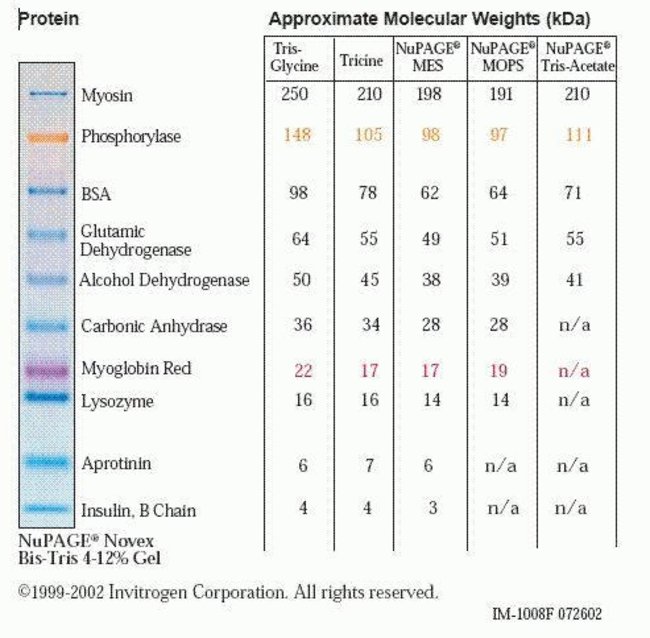
Find additional tips, troubleshooting help, and resources within our Protein Standards and Ladders Support Center.
SeeBlue Plus2 Pre-Stained Standard includes 10 recombinant proteins, that resolve in the range of 4 to 250 kDa depending upon the buffer system being used. Please note that this pre-stained protein standard is not designed for quantitation and we do not recommend that you use this standard in the determination of protein concentration.
Estimations of the protein concentration for SeeBlue Plus2 is given below. However, these values are an approximation and should not be used to quantify samples.
Band per 10 µL: Myosin - 2.20 µg; Phosphorylase-b no estimation is available; BSA - 0.75 µg; GDH - 1.25 µg; Alcohol Dehydrogenase - 0.80 µg; Carbonic Anhydrase- 0.90 µg; Myoglobin (blue)* no estimation available; Lysozyme - 2.50 µg; Aprotinin - 1.80 µg and Insulin - 2.50 µg.
Find additional tips, troubleshooting help, and resources within our Protein Electrophoresis and Western Blotting Support Center.
The fading is most likely due to detergent in the western blocking/washing solutions that can remove some of the proteins from the membrane. The dye itself will not wash off of the proteins because it is covalently bound. We have found that smaller pore size membranes retain the proteins better during blocking and wash procedures, and hence recommend use of 0.2 µm instead of 0.45 µm membranes for best resolution and protein retention. After transfer, it is a good idea to circle the pre-stained bands with a pencil on the membrane, so band positions can be identified after blocking and processing.
Find additional tips, troubleshooting help, and resources within our Protein Assays and Analysis Support Center.