Search Thermo Fisher Scientific


SDS

Citations & References (5)
Invitrogen™
SimplyBlue™ SafeStain
SimplyBlue SafeStain is a ready-to-use, fast, sensitive, and safe Coomassie G-250 stain for visualizing protein bands on polyacrylamide gels and on dry PVDF membranes.
Have Questions?
Change view

| Catalog Number | Quantity |
|---|---|
| LC6060 | 1 L |
| LC6065 | 3.5 L |
Catalog number LC6060
Price (USD)
231.00
Each
-
Quantity:
1 L
Price (USD)
231.00
Each
SimplyBlue SafeStain is a ready-to-use, fast, sensitive, and safe Coomassie G-250 stain for visualizing protein bands on polyacrylamide gels and on dry PVDF membranes. SimplyBlue SafeStain is completely non-hazardous and does not require methanol or acetic acid fixatives or destains. Risk of hazardous exposure and unpleasant odors are eliminated, creating a healthier lab environment.
Coomassie G-250 stain for visualizing protein bands on polyacrylamide gels and on dry PVDF membranes. SimplyBlue SafeStain is completely non-hazardous and does not require methanol or acetic acid fixatives or destains. Risk of hazardous exposure and unpleasant odors are eliminated, creating a healthier lab environment.
Features include:
• An easy-to-perform protocol that can be completed in less than three hours. For added speed, a simple microwave procedure can be completed in 12 minutes
• Destaining not required but may be performed with deionized water to achieve maximum sensitivity, especially when performing downstream analyses such as mass spectrometry, or when a crystal-clear background is required
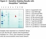
• Sensitive—detect down to 7 ng of protein
• Fast—detect 1 μg of protein in as little as five minutes after staining
• Convenient—save valuable time by skipping reagent preparation with this ready-to-use formulation
• Non-hazardous—save money and hassle associated with hazardous shipping and disposal of hazardous waste
Features include:
• An easy-to-perform protocol that can be completed in less than three hours. For added speed, a simple microwave procedure can be completed in 12 minutes
• Destaining not required but may be performed with deionized water to achieve maximum sensitivity, especially when performing downstream analyses such as mass spectrometry, or when a crystal-clear background is required
• Sensitive—detect down to 7 ng of protein
• Fast—detect 1 μg of protein in as little as five minutes after staining
• Convenient—save valuable time by skipping reagent preparation with this ready-to-use formulation
• Non-hazardous—save money and hassle associated with hazardous shipping and disposal of hazardous waste
For Research Use Only. Not for use in diagnostic procedures.
Specifications
Detection LocationIn-Gel Detection
Detection MethodColorimetric
Green FeaturesLess hazardous
Product LineSimplyBlue™
Product TypeSafe Protein Stain
Quantity1 L
Shelf Life6 Months
Target MoleculeProtein
ColorBlue
Label or DyeCoomassie
Unit SizeEach
Contents & Storage
SimplyBlue™ SafeStain is supplied as a 1X ready-to-use staining reagent. Store at room temperature. Guaranteed stable for 6 months when properly stored. There are no HazMat charges associated with this product.

Introducing iBlot 3 Western Blot Transfer System
Featuring higher throughput and built-in cooling for consistent protein transfer
Learn more ›
Have questions about this product? Ask our AI assisted search.
This is an AI-powered search and may not always get things right. You can help us make it better with a thumbs up or down on individual answers or by selecting the “Give feedback" button. Your search history and customer login information may be retained by Thermo Fisher and processed in accordance with our
Privacy Notice.
Figures

Sensitive staining with SimplyBlue™ SafeStain.

Rapid staining with SimplyBlue™ SafeStain.
Customers who viewed this item also viewed
Documents & Downloads
Certificates
Search by lot number or partial lot number
0 results displayed, search above for a specific certificate
Safety Data Sheets
SDSFrequently asked questions (FAQs)
It is not recommended because the background will be too high. Better alternatives include:
1) Invitrogen Reversible Membrane Protein Stain Kit (Cat. No. IB7710).
2) Coomassie (non-colloidal) staining: stain in 0.1% Coomassie Blue R-250 in 50% methanol for 5 min and destain with several changes of 50% methanol and 10% acetic acid. Rinse with several changes of water, air dry and store for up to 12 months at -20°C. Sensitivity is approximately at the 50-100 ng level.
3) Use SimplyBlue SafeStain (Cat. No. LC6060). The SimplyBlue SafeStain manual has the protocol for staining PVDF membranes, but it is not recommended for nitrocellulose because of high background.
4) Amido Black: same as Coomassie but less sensitive.
5) Ponceau S: same as Coomassie but less sensitive.
6) UV transillumination: place membrane on filter paper after blot is finished and allow to dry at room temperature for about 10 min. Rewet in 20% methanol and view the blot in front of white light while it is still wet; the bands will look more translucent than the membrane. If the bands disappear as the membranes dries, rewet again.
Find additional tips, troubleshooting help, and resources within our Protein Electrophoresis and Western Blotting Support Center.
Coomassie G-250 will give a sharp dye front with both NuPAGE MES and MOPS Running Buffers and is therefore used as the tracking dye in the NuPAGE LDS Sample Buffer.
Bromophenol blue runs more slowly than some peptides with the NuPAGE MES Running Buffer system.
Coomassie G-250 migrates much closer to the moving ion front than bromophenol blue, ensuring that small peptides will not be run too far (e.g., off the gel).
Find additional tips, troubleshooting help, and resources within our Protein Electrophoresis and Western Blotting Support Center.
The Colloidal Blue Staining Kit (Cat. No. LC6025) is best for quantitation by densitometry. You can also use SimplyBlue SafeStain for this application.
The great advantage of SimplyBlue SafeStain is that it is very easy to use and safe.
Find additional tips, troubleshooting help, and resources within our Protein Assays and Analysis Support Center.
Check the cap on the bottle. If the bottles are not tightly sealed, the alcohol can evaporate from the stain causing substantial decrease in stain sensitivity.
Find additional tips, troubleshooting help, and resources within our Protein Assays and Analysis Support Center.
After staining with SimplyBlue SafeStain, use deionized water for the less strongly retained protein bands on the PVDF membrane.
Increasing methanol or ethanol concentrations up to 70% should destain any remaining bands. You can leave the membrane in the destain indefinitely.
Find additional tips, troubleshooting help, and resources within our Protein Assays and Analysis Support Center.
Citations & References (5)
Search citations by name, author, journal title or abstract text
Citations & References
Abstract
Enzymatic hydrolysis of pyridoxine-5'-beta-D-glucoside is catalyzed by intestinal lactase-phlorizin hydrolase.
Journal:J Biol Chem
PubMed ID:12023280
'An obligatory step in the mammalian nutritional utilization of pyridoxine-5''-beta-D-glucoside (PNG) is the intestinal hydrolysis of its beta-glucosidic bond that releases pyridoxine (PN). This laboratory previously reported the purification and partial characterization of a novel cytosolic enzyme, designated PNG hydrolase, which hydrolyzed PNG. An investigation of the subcellular distribution of
2F3 monoclonal antibody recognizes the O26 O-antigen moiety of the lipopolysaccharide of enterohemorrhagic Escherichia coli strain 4276.
Journal:Clin Diagn Lab Immunol
PubMed ID:15138178
'Enterohemorrhagic Escherichia coli (EHEC) and enteropathogenic E. coli (EPEC) organisms are groups of pathogenic strains whose infections are characterized by a typical lesion of enterocyte attachment and effacement. They are involved in enteric diseases both in humans and in animals, and EHEC strains can be responsible for hemolytic uremic syndrome
Selective fluorescent labeling of S-nitrosothiols (S-FLOS): a novel method for studying S-nitrosation.
Journal:Nitric Oxide
PubMed ID:18706513
'Protein S-nitrosation is a reversible post-translation modification critical for redox-sensitive cell signaling that is typically studied using the Biotin Switch method. This method and subsequent modifications usually require avidin binding or Western blot analysis to detect biotin labeled proteins. We describe here a modification of the Biotin Switch assay that
Vascular endothelial growth factor-C and C-C chemokine receptor 7 in tumor cell-lymphatic cross-talk promote invasive phenotype.
Journal:Cancer Res
PubMed ID:19118020
'Most carcinomas spread to distant sites through lymphatic vessels. Several preclinical and clinical studies have shown a positive correlation between the incidence of lymph node metastasis and secretion of the lymphatic growth factor vascular endothelial growth factor-C (VEGF-C) by tumor cells, suggesting tumor lymphangiogenesis as an escape mechanism. However, recent
Yeast Expression and NMR Analysis of the Extracellular Domain of Muscle Nicotinic Acetylcholine Receptor alpha Subunit.
Journal:J Biol Chem
PubMed ID:11812776
The alpha subunit of the nicotinic acetylcholine receptor (AChR) from Torpedo electric organ and mammalian muscle contains high affinity binding sites for alpha-bungarotoxin and for autoimmune antibodies in sera of patients with myasthenia gravis. To obtain sufficient materials for structural studies of the receptor-ligand complexes, we have expressed part of
5 total citations