Search Thermo Fisher Scientific

Materials Science
Metals Research
Characterization of metals, ranging from large surfaces to inclusions and precipitates.
bf5a- Apps/Techniques/Products/Resources/Contact Us
Modern cutting-edge metals are increasingly engineered at the nanoscale to enhance their durability, reliability, and cost. Even traditional processes are now augmented with microscopic inspection to determine the resulting material’s elemental and structural composition.
In particular, the effective production of metals requires precise control of inclusions and precipitates. Depending on their consistency and distribution, these can either strengthen the material or act as contaminants, greatly impacting quality and lifetime. These microscopic properties can include;
- Nano-precipitates formed during rolling, annealing or hot pressing
- Nanoscale morphological changes (dislocations, crack initiation sites)
- Grain boundaries
- Oxide inclusions that cause casting interruptions in steelmaking
Historically, researchers have used optical microscopy to rate the size and number of inclusions, but this method does not provide any elemental information. Even optical emission spectroscopy, which can determine the elemental ratios of inclusions, does not accurately characterize the shape and composition of individual inclusions. Electron microscopy techniques have also been used for metal analysis, with scanning electron microscopy (SEM) capable of visualizing larger oxide inclusions, whereas transmission electron microscopy (TEM) is generally required to study features smaller than 100 nm. TEM analysis, however, has previously required manual particle counting and analysis, limiting the amount of data that could be collected to several dozen particles per day.

Stainless steel medical device sample generated with PFIB milling, with total dimensions of 55 x 70 μm. The red box indicates the amount of area that could be prepared in the same amount of time with a typical gallium FIB.
Thermo Fisher Scientific provides a range of electron microscopy solutions that make metal analysis not only more informative but also far more rapid. Thanks to our unique automation capabilities, a thorough overview of the elemental and structural composition of hundreds, if not thousands, of precipitates is possible in a manner of hours, as compared to the few dozen that would be found in a day of manual analysis. Not only is statistical information on the bulk available, but individual precipitates can also be seen with high detail, providing a multi-scale overview of the metal.
Our robust, automated instruments can perform a variety of critical tasks including:
- Nanoparticle counting, particularly useful for steel and aluminum production where light weighting is a top priority
- High throughput chemical analysis with energy-dispersive X-ray spectroscopy (EDS) mapping
- Instantly showing composition gradients across the sample surface with scanning electron microscopy (SEM) via ChemiSEM technology
- Rapidly preparing large area transmission electron microscopy samples or 3D volumes with plasma focused ion beam (PFIB) milling

3D microstructural information provided by electron backscatter diffraction (EBSD) of a zirconium alloy sample reconstructed from 400 slices. Sample courtesy of the University of Manchester.
SEM images

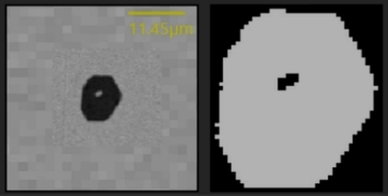
Low-carbon, aluminum-killed steel sample observed by Phenom ParticleX Steel Desktop SEM. Backscattered imaging shows a cluster of several micron-sized alumina inclusions.

Titanium-stabilized, ultra-low-carbon steel sample observed by Phenom ParticleX Steel Desktop SEM. Backscattered imaging shows cubic titanium nitride precipitating on top of an oxide inclusion.

Calcium-treated steel sample observed by Phenom ParticleX Steel Desktop SEM. Backscattered imaging shows MgO.Al2O3 spinel phase (dark) forming inside a calcium aluminate inclusion.

Calcium-treated steel was scanned by a Phenom ParticleX Steel Desktop SEM over 60 mm2 to characterize non-metallic inclusions. This ternary diagram reveals the composition distribution of calcium aluminates and calcium sulfides in this steel alloy.

Calcium-treated steel was scanned by a Phenom ParticleX Steel Desktop SEM over 60 mm2 to characterize non-metallic inclusions. This particle classification table shows the count and average composition of over 1,500 inclusions that were identified.

Calcium-treated steel sample observed by a Phenom ParticleX Steel Desktop SEM. Backscattered imaging (left) and EDS mapping show a compound calcium sulfide and calcium aluminate inclusion. On the EDS maps, calcium is shown in yellow and aluminum in blue.
 and small (< 1 μm) titanium rich particles. Sample courtesy of GKN Aerospace.")
Forged steel cylinder polished section observed by Phenom ParticleX Steel. Backscattered imaging reveals the spatial distribution of large (> 5 μm) and small (< 1 μm) titanium rich particles. Sample courtesy of GKN Aerospace.

Forged steel cylinder polished section was scanned by ParticleX Steel over 50 mm2 to characterize non-metallic inclusions. This ternary diagram reveals the size and composition distribution of titanium sulfides and titanium nitrides in this steel alloy. Sample courtesy of GKN Aerospace.

Forged steel cylinder surface observed by Axia ChemiSEM. This area has been machined which exposed large non-metallic phases. ChemiSEM EDS mapping confirmed that the particles are titanium rich. Sample courtesy of GKN Aerospace.

Friction stir welded aerospace aluminum alloy was scanned by ParticleX Steel over 568 mm2 to characterize particles brighter than the base metal. This Fe-Mn-Cu ternary diagram shows the chemical distribution of 65k bright phase particles. Sample courtesy of GKN Aerospace.

Friction stir welded aerospace aluminum alloy was scanned by ParticleX Steel over 568 mm2 to characterize particles brighter than the base metal. This particle table shows the majority of the 65k particles contain a portion of iron, manganese and copper. Sample courtesy of GKN Aerospace.

Friction stir welded aerospace aluminum alloy was scanned by ParticleX Steel to characterize heavy metal particles. Backscattered electron image reveals bright phase particles which have a higher average atomic weight than the base metal. Sample courtesy of GKN Aerospace.
XPS images

Forged steel cylinder surface analyzed by the Thermo Scientific Nexsa G2 Surface Analysis System. Optical micrograph reveals some of the machining detail on this heat treated sample. Sample courtesy of GKN Aerospace.

Forged steel cylinder surface analyzed by XPS with depth profiling. It reveals the passivated layer of chromium oxide with the unoxidized steel beneath. Sample courtesy of GKN Aerospace.
TEM images
 and zirconium (red), msd-em, Metals research, 280x200")
 and zirconium (red)")
 and zirconium (red)")
Precipitates containing copper (green) and zirconium (red) in a friction-stir-welded Al-Cu-Li alloy were analyzed with a Talos F200X (S)TEM and Automated Particle Workflow (APW). The three regions represent the base metal (left), the heat-affected zone (middle), and the stirred zone (right).


Precipitates of niobium carbide in a high-strength, low-alloy steel were analyzed with a Talos F200X (S)TEM and Automated Particle Workflow (APW). The two regions represent different locations on the same coil, where the steel with finer precipitates (average 9 nm, left) yielded a higher strength than the steel with larger precipitates (average 12 nm, right).
 HAADF STEM image, b) zinc EDS map, c) zinc particle segmentation. Sample courtesy of University of Manchester and University of Trento.")
Talos F200X S/TEM analysis of Aluminum 7075 aerospace alloy showing a) HAADF STEM image, b) zinc EDS map, c) zinc particle segmentation. Sample courtesy of University of Manchester and University of Trento.
 magnesium particle segmentation, b) zinc particle segmentation, and c) Co-Located magnesium and zinc compounds shown in orange. Sample courtesy of University of Manchester and University of Trento.")
Talos F200X S/TEM analysis of Aluminum 7075 aerospace alloy showing a) magnesium particle segmentation, b) zinc particle segmentation, and c) Co-Located magnesium and zinc compounds shown in orange. Sample courtesy of University of Manchester and University of Trento.
 HAADF STEM image and b) corresponding APW particle map of magnesium and zinc compounds. Sample courtesy of University of Manchester and University of Trento.")
Talos F200X S/TEM analysis of Aluminum 7075 aerospace alloy showing a) HAADF STEM image and b) corresponding APW particle map of magnesium and zinc compounds. Sample courtesy of University of Manchester and University of Trento.

Talos F200X S/TEM analysis of high strength low alloy steel carbon replica. HAADF STEM image shows precipitates and grain boundaries as a lighter color on a dark background. Sample courtesy of OCAS.

Talos F200X S/TEM analysis via Automated Particle Workflow of high strength low alloy steel carbon replica. Zoom area of the segmented particle map shows titanium in gold, niobium in pink and where they overlap is orange. Sample courtesy of OCAS.
 size distribution, and segmented particle map shows titanium in gold, niobium in pink and where they overlap is orange. Sample courtesy of OCAS.")
Talos F200X S/TEM analysis via Automated Particle Workflow of high strength low alloy steel carbon replica. Chart shows compound particle (TiN + NbC) size distribution, and segmented particle map shows titanium in gold, niobium in pink and where they overlap is orange. Sample courtesy of OCAS.
 HAADF STEM image of a precipitate in the TEM lamella, and b) manual EDS mapping of the complex precipitate which contains a majority of MnSi oxide at the center and precipitates of Cu and NbN on the perimeter. Sample courtesy of University of Connecticut.")
Talos F200X S/TEM analysis of additively manufactured 17-4 PH stainless steel showing: a) HAADF STEM image of a precipitate in the TEM lamella, and b) manual EDS mapping of the complex precipitate which contains a majority of MnSi oxide at the center and precipitates of Cu and NbN on the perimeter. Sample courtesy of University of Connecticut.
 and Niobium (red) or Avizo particle quantification of silicon particles. Sample courtesy of University of Connecticut")
High resolution APW analysis showing EDS maps of Silicon (green) and Niobium (red) or Avizo particle quantification of silicon particles. Sample courtesy of University of Connecticut
 and niobium (blue). Sample courtesy of University of Connecticut.")
High resolution APW analysis showing compounds precipitates of silicon (yellow) and niobium (blue). Sample courtesy of University of Connecticut.
SEM videos
Phenom ParticleX Steel Desktop SEM inclusion analysis short demonstration.
ParticleX Steel Desktop SEM - Workflow introduction.
Axia ChemiSEM provides high-quality imaging of steel samples to aid in the production of high-value steels.
Axia ChemiSEM identifies composition on-the-fly
TEM videos

Aluminum 2099 alloy lamella characterization of Cu and Zr precipitates by APW
HSLA steel lamella characterization of Nb precipitates by Automated Particle Workflow (APW).
3D EDS TEM tomography of precipitates in an AlMgSi alloy.
High resolution APW showing complex features in additively manufactured stainless steel.
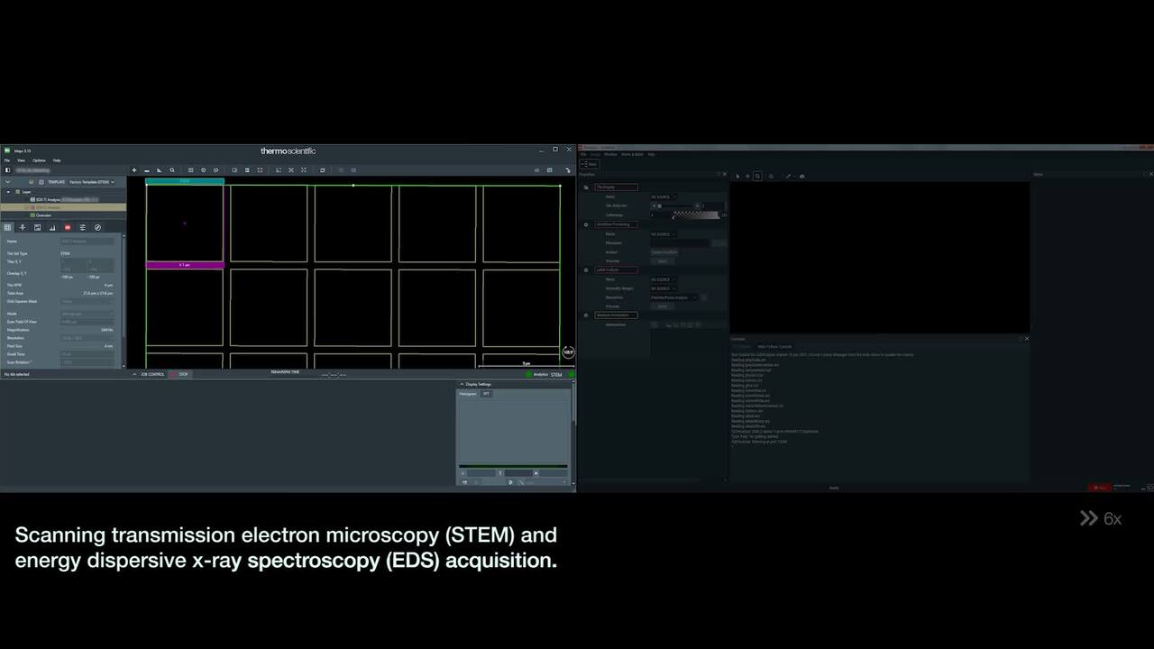
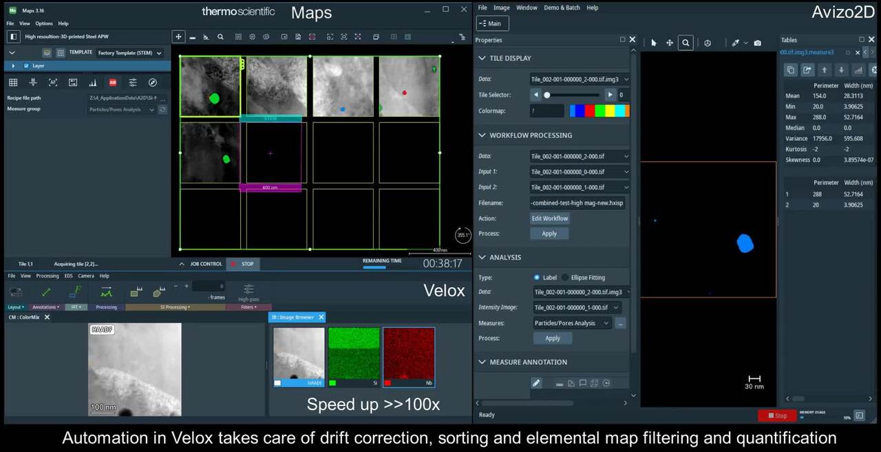
Maps and Avizo2D recordings (left and right) running side by side during an acquisition.
Webinars


Webinar: Nanoparticle Characterization by Automated TEM.
Webinar: Correlative Microscopy for Aerospace and Defense Industries


")

Documents
SEM documents
TEM documents
TEM Articles
Nanoscale origins of the oriented precipitation of Ti3Al in Ti\\Al systems
Hao Wu, Guohua Fan, Lin Geng, Xiping Cui, Meng Huang
Effect of heat treatments on microstructural evolution of additively manufactured and wrought 17-4PH stainless steel
Yu Sun, Rainer J. Hebert, Mark Aindow
Coherency strains of H-phase precipitates and their influence on functional properties of nickel-titanium-hafnium shape memory alloys
Behnam Amin-Ahmadi,⁎, Joseph G. Pauza, Ali Shamimi, Tom W. Duerig, Ronald D. Noebe, Aaron P. Stebner
Effect of laser scan length on the microstructure of additively manufactured 17-4PH stainless steel thin-walled parts
Yu Sun, Rainer J. Hebert, Mark Aindow
Non-metallic inclusions in 17-4PH stainless steel parts produced by selective laser melting
Yu Sun, Rainer J. Hebert, Mark Aindow
FIB-SEM Articles
Joachim Mayer, RWTH Aachen
“Formation of White Etching Areas in SAE 52100 Bearing Steel under Rolling Contact Fatigue − Influence of Diffusible Hydrogen”
M. Oezel, A. Schwedt, T. Janitzky, R. Kelley, C.Bouchet-Marquis, L. Pullan, C. Broeckmann, J. Mayer
Wear, Volumes 414-415, November 2018, Pages 352-365.
Philip Withers, University of Manchester
“Industrial Gear Oils: Tribological Performance and Subsurface Changes”
Aduragbemi Adebogun, Robert Hudson, Angela Breakspear, Chris Warrens, Ali Gholinia, Allan Matthews, Philip Withers Tribology Letters (2018) 66:65.
Jun Tan, Shenyang National Laboratory for Materials Science
“Insight into atmospheric pitting corrosion of carbon steel via a dual-beam FIB/SEM system associated with high-resolution TEM”
Corrosion Science 152 (2019) 226–233.
Yu-Lung Chiu, University of Birmingham
“Micro-tensile strength of a welded turbine disc superalloy”
K.M. Oluwasegun, C.Cooper, Y.L.Chiu, I.P.Jones, H.Y.Li, G.Baxter
Materials Science & Engineering A 596 (2014) 229–235.
Chris Pistorius, Carnegie Mellon University
“Application of Plasma FIB to Analyze a Single Oxide Inclusion in Steel”
D. Kumar, N.T. Nuhfer, M.E. Ferreira and P.C. Pistorius
Metallurgical and Materials Transactions B, Volume 50B, June 2019, Pages 1124-1127.
SEM images
Low-carbon, aluminum-killed steel sample observed by Phenom ParticleX Steel Desktop SEM. Backscattered imaging shows a cluster of several micron-sized alumina inclusions.
Titanium-stabilized, ultra-low-carbon steel sample observed by Phenom ParticleX Steel Desktop SEM. Backscattered imaging shows cubic titanium nitride precipitating on top of an oxide inclusion.
Calcium-treated steel sample observed by Phenom ParticleX Steel Desktop SEM. Backscattered imaging shows MgO.Al2O3 spinel phase (dark) forming inside a calcium aluminate inclusion.
Calcium-treated steel was scanned by a Phenom ParticleX Steel Desktop SEM over 60 mm2 to characterize non-metallic inclusions. This ternary diagram reveals the composition distribution of calcium aluminates and calcium sulfides in this steel alloy.
Calcium-treated steel was scanned by a Phenom ParticleX Steel Desktop SEM over 60 mm2 to characterize non-metallic inclusions. This particle classification table shows the count and average composition of over 1,500 inclusions that were identified.
Calcium-treated steel sample observed by a Phenom ParticleX Steel Desktop SEM. Backscattered imaging (left) and EDS mapping show a compound calcium sulfide and calcium aluminate inclusion. On the EDS maps, calcium is shown in yellow and aluminum in blue.
Forged steel cylinder polished section observed by Phenom ParticleX Steel. Backscattered imaging reveals the spatial distribution of large (> 5 μm) and small (< 1 μm) titanium rich particles. Sample courtesy of GKN Aerospace.
Forged steel cylinder polished section was scanned by ParticleX Steel over 50 mm2 to characterize non-metallic inclusions. This ternary diagram reveals the size and composition distribution of titanium sulfides and titanium nitrides in this steel alloy. Sample courtesy of GKN Aerospace.
Forged steel cylinder surface observed by Axia ChemiSEM. This area has been machined which exposed large non-metallic phases. ChemiSEM EDS mapping confirmed that the particles are titanium rich. Sample courtesy of GKN Aerospace.
Friction stir welded aerospace aluminum alloy was scanned by ParticleX Steel over 568 mm2 to characterize particles brighter than the base metal. This Fe-Mn-Cu ternary diagram shows the chemical distribution of 65k bright phase particles. Sample courtesy of GKN Aerospace.
Friction stir welded aerospace aluminum alloy was scanned by ParticleX Steel over 568 mm2 to characterize particles brighter than the base metal. This particle table shows the majority of the 65k particles contain a portion of iron, manganese and copper. Sample courtesy of GKN Aerospace.
Friction stir welded aerospace aluminum alloy was scanned by ParticleX Steel to characterize heavy metal particles. Backscattered electron image reveals bright phase particles which have a higher average atomic weight than the base metal. Sample courtesy of GKN Aerospace.
XPS images
Forged steel cylinder surface analyzed by the Thermo Scientific Nexsa G2 Surface Analysis System. Optical micrograph reveals some of the machining detail on this heat treated sample. Sample courtesy of GKN Aerospace.
Forged steel cylinder surface analyzed by XPS with depth profiling. It reveals the passivated layer of chromium oxide with the unoxidized steel beneath. Sample courtesy of GKN Aerospace.
TEM images
Precipitates containing copper (green) and zirconium (red) in a friction-stir-welded Al-Cu-Li alloy were analyzed with a Talos F200X (S)TEM and Automated Particle Workflow (APW). The three regions represent the base metal (left), the heat-affected zone (middle), and the stirred zone (right).
Precipitates of niobium carbide in a high-strength, low-alloy steel were analyzed with a Talos F200X (S)TEM and Automated Particle Workflow (APW). The two regions represent different locations on the same coil, where the steel with finer precipitates (average 9 nm, left) yielded a higher strength than the steel with larger precipitates (average 12 nm, right).
Talos F200X S/TEM analysis of Aluminum 7075 aerospace alloy showing a) HAADF STEM image, b) zinc EDS map, c) zinc particle segmentation. Sample courtesy of University of Manchester and University of Trento.
Talos F200X S/TEM analysis of Aluminum 7075 aerospace alloy showing a) magnesium particle segmentation, b) zinc particle segmentation, and c) Co-Located magnesium and zinc compounds shown in orange. Sample courtesy of University of Manchester and University of Trento.
Talos F200X S/TEM analysis of Aluminum 7075 aerospace alloy showing a) HAADF STEM image and b) corresponding APW particle map of magnesium and zinc compounds. Sample courtesy of University of Manchester and University of Trento.
Talos F200X S/TEM analysis of high strength low alloy steel carbon replica. HAADF STEM image shows precipitates and grain boundaries as a lighter color on a dark background. Sample courtesy of OCAS.
Talos F200X S/TEM analysis via Automated Particle Workflow of high strength low alloy steel carbon replica. Zoom area of the segmented particle map shows titanium in gold, niobium in pink and where they overlap is orange. Sample courtesy of OCAS.
Talos F200X S/TEM analysis via Automated Particle Workflow of high strength low alloy steel carbon replica. Chart shows compound particle (TiN + NbC) size distribution, and segmented particle map shows titanium in gold, niobium in pink and where they overlap is orange. Sample courtesy of OCAS.
Talos F200X S/TEM analysis of additively manufactured 17-4 PH stainless steel showing: a) HAADF STEM image of a precipitate in the TEM lamella, and b) manual EDS mapping of the complex precipitate which contains a majority of MnSi oxide at the center and precipitates of Cu and NbN on the perimeter. Sample courtesy of University of Connecticut.
High resolution APW analysis showing EDS maps of Silicon (green) and Niobium (red) or Avizo particle quantification of silicon particles. Sample courtesy of University of Connecticut
High resolution APW analysis showing compounds precipitates of silicon (yellow) and niobium (blue). Sample courtesy of University of Connecticut.
SEM videos
Phenom ParticleX Steel Desktop SEM inclusion analysis short demonstration.
ParticleX Steel Desktop SEM - Workflow introduction.
Axia ChemiSEM provides high-quality imaging of steel samples to aid in the production of high-value steels.
Axia ChemiSEM identifies composition on-the-fly
TEM videos
Aluminum 2099 alloy lamella characterization of Cu and Zr precipitates by APW
HSLA steel lamella characterization of Nb precipitates by Automated Particle Workflow (APW).
3D EDS TEM tomography of precipitates in an AlMgSi alloy.
High resolution APW showing complex features in additively manufactured stainless steel.
Maps and Avizo2D recordings (left and right) running side by side during an acquisition.
Webinars
Webinar: Nanoparticle Characterization by Automated TEM.
Webinar: Correlative Microscopy for Aerospace and Defense Industries
Documents
SEM documents
TEM documents
TEM Articles
Nanoscale origins of the oriented precipitation of Ti3Al in Ti\\Al systems
Hao Wu, Guohua Fan, Lin Geng, Xiping Cui, Meng Huang
Effect of heat treatments on microstructural evolution of additively manufactured and wrought 17-4PH stainless steel
Yu Sun, Rainer J. Hebert, Mark Aindow
Coherency strains of H-phase precipitates and their influence on functional properties of nickel-titanium-hafnium shape memory alloys
Behnam Amin-Ahmadi,⁎, Joseph G. Pauza, Ali Shamimi, Tom W. Duerig, Ronald D. Noebe, Aaron P. Stebner
Effect of laser scan length on the microstructure of additively manufactured 17-4PH stainless steel thin-walled parts
Yu Sun, Rainer J. Hebert, Mark Aindow
Non-metallic inclusions in 17-4PH stainless steel parts produced by selective laser melting
Yu Sun, Rainer J. Hebert, Mark Aindow
FIB-SEM Articles
Joachim Mayer, RWTH Aachen
“Formation of White Etching Areas in SAE 52100 Bearing Steel under Rolling Contact Fatigue − Influence of Diffusible Hydrogen”
M. Oezel, A. Schwedt, T. Janitzky, R. Kelley, C.Bouchet-Marquis, L. Pullan, C. Broeckmann, J. Mayer
Wear, Volumes 414-415, November 2018, Pages 352-365.
Philip Withers, University of Manchester
“Industrial Gear Oils: Tribological Performance and Subsurface Changes”
Aduragbemi Adebogun, Robert Hudson, Angela Breakspear, Chris Warrens, Ali Gholinia, Allan Matthews, Philip Withers Tribology Letters (2018) 66:65.
Jun Tan, Shenyang National Laboratory for Materials Science
“Insight into atmospheric pitting corrosion of carbon steel via a dual-beam FIB/SEM system associated with high-resolution TEM”
Corrosion Science 152 (2019) 226–233.
Yu-Lung Chiu, University of Birmingham
“Micro-tensile strength of a welded turbine disc superalloy”
K.M. Oluwasegun, C.Cooper, Y.L.Chiu, I.P.Jones, H.Y.Li, G.Baxter
Materials Science & Engineering A 596 (2014) 229–235.
Chris Pistorius, Carnegie Mellon University
“Application of Plasma FIB to Analyze a Single Oxide Inclusion in Steel”
D. Kumar, N.T. Nuhfer, M.E. Ferreira and P.C. Pistorius
Metallurgical and Materials Transactions B, Volume 50B, June 2019, Pages 1124-1127.

Process control using electron microscopy
Modern industry demands high throughput with superior quality, a balance that is maintained through robust process control. SEM and TEM tools with dedicated automation software provide rapid, multi-scale information for process monitoring and improvement.

Quality control and failure analysis
Quality control and assurance are essential in modern industry. We offer a range of EM and spectroscopy tools for multi-scale and multi-modal analysis of defects, allowing you to make reliable and informed decisions for process control and improvement.

Fundamental Materials Research
Novel materials are investigated at increasingly smaller scales for maximum control of their physical and chemical properties. Electron microscopy provides researchers with key insight into a wide variety of material characteristics at the micro- to nano-scale.

Technical Cleanliness
More than ever, modern manufacturing necessitates reliable, quality components. With scanning electron microscopy, parts cleanliness analysis can be brought inhouse, providing you with a broad range of analytical data and shortening your production cycle.
Style Sheet for Komodo Tabs

(S)TEM Sample Preparation
DualBeam microscopes enable the preparation of high-quality, ultra-thin samples for (S)TEM analysis. Thanks to advanced automation, users with any experience level can obtain expert-level results for a wide range of materials.

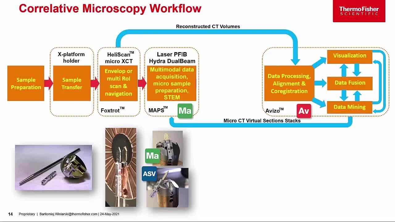
3D Materials Characterization
Development of materials often requires multi-scale 3D characterization. DualBeam instruments enable serial sectioning of large volumes and subsequent SEM imaging at nanometer scale, which can be processed into high-quality 3D reconstructions of the sample.

Energy Dispersive Spectroscopy
Energy dispersive spectroscopy (EDS) collects detailed elemental information along with electron microscopy images, providing critical compositional context for EM observations. With EDS, chemical composition can be determined from quick, holistic surface scans down to individual atoms.
_Technique_800x375_144DPI.jpg)
EDS Elemental Analysis
Thermo Scientific Phenom Elemental Mapping Software provides fast and reliable information on the distribution of chemical elements within a sample.
_Technique_800x375_144DPI.jpg)
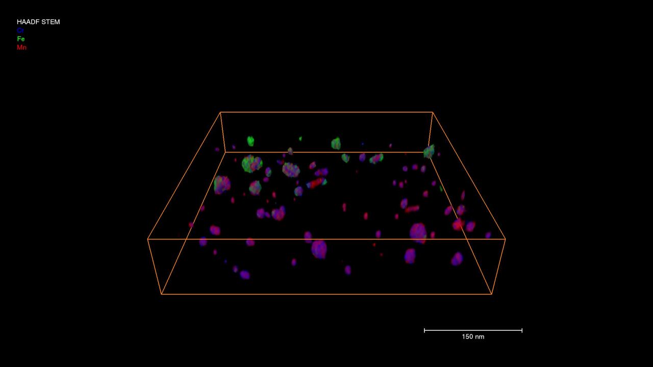
3D EDS Tomography
Modern materials research is increasingly reliant on nanoscale analysis in three dimensions. 3D characterization, including compositional data for full chemical and structural context, is possible with 3D EM and energy dispersive X-ray spectroscopy.

EDS Analysis with ChemiSEM Technology
Energy dispersive X-ray spectroscopy for materials characterization.

Cross-sectioning
Cross sectioning provides extra insight by revealing sub-surface information. DualBeam instruments feature superior focused ion beam columns for high-quality cross sectioning. With automation, unattended high-throughput processing of samples is possible.

In Situ experimentation
Direct, real-time observation of microstructural changes with electron microscopy is necessary to understand the underlying principles of dynamic processes such as recrystallization, grain growth, and phase transformation during heating, cooling, and wetting.

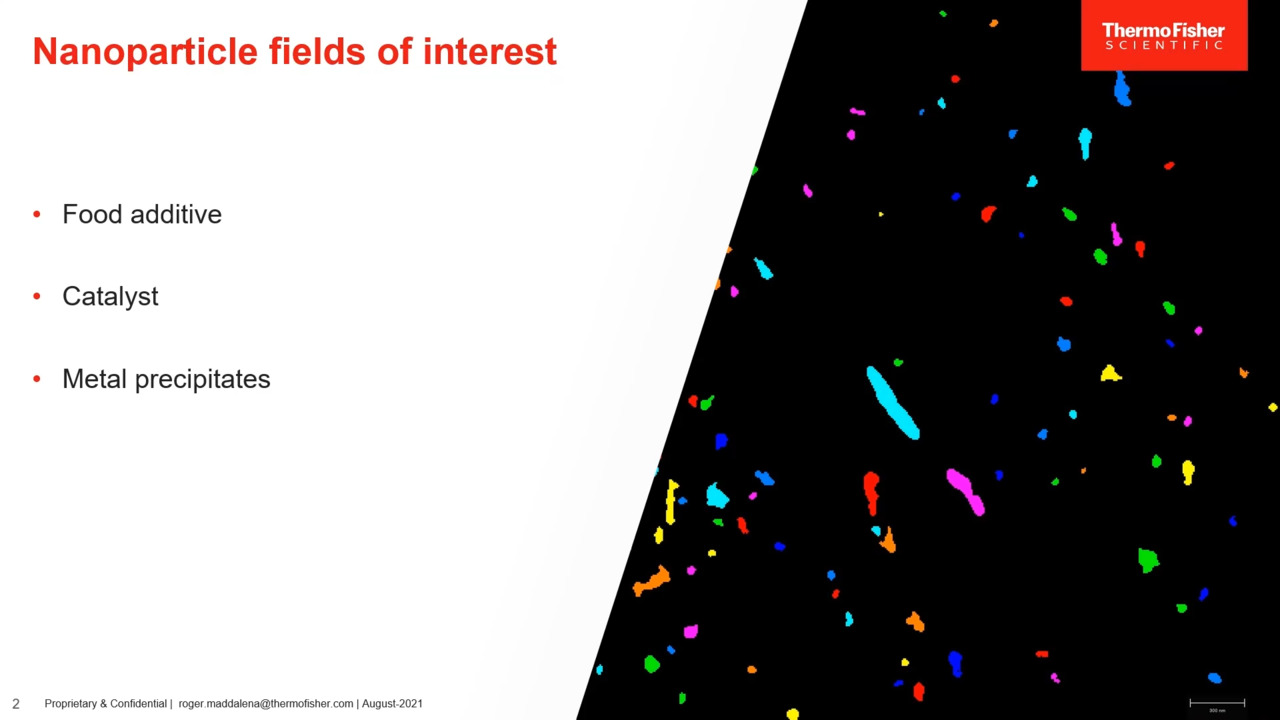
Particle analysis
Particle analysis plays a vital role in nanomaterials research and quality control. The nanometer-scale resolution and superior imaging of electron microscopy can be combined with specialized software for rapid characterization of powders and particles.

X-Ray Photoelectron Spectroscopy
X-ray photoelectron spectroscopy (XPS) enables surface analysis, providing elemental composition as well as the chemical and electronic state of the top 10 nm of a material. With depth profiling, XPS analysis extends to compositional insight of layers.

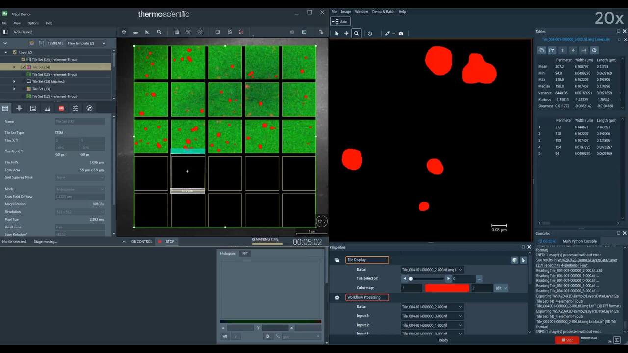
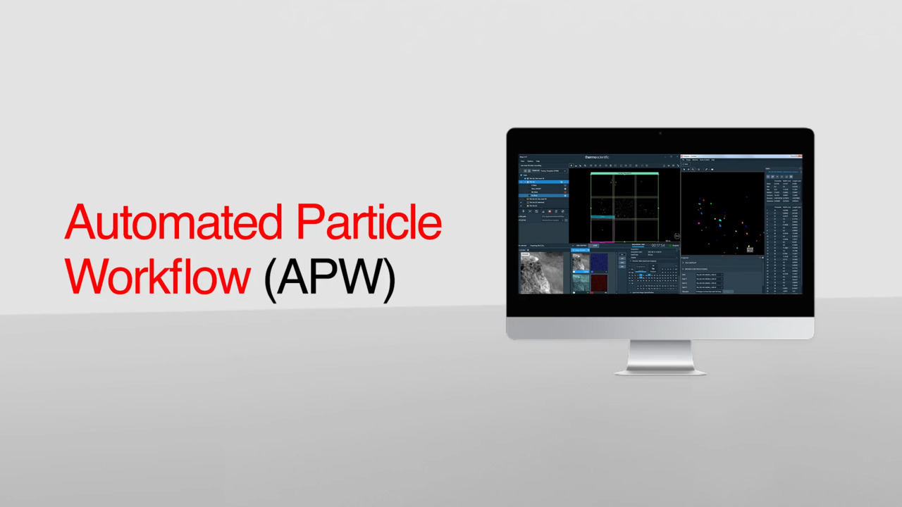
The Automated NanoParticle Workflow (APW) is a transmission electron microscope workflow for nanoparticle analysis, offering large area, high resolution imaging and data acquisition at the nanoscale, with on-the-fly processing.
(S)TEM Sample Preparation
DualBeam microscopes enable the preparation of high-quality, ultra-thin samples for (S)TEM analysis. Thanks to advanced automation, users with any experience level can obtain expert-level results for a wide range of materials.
3D Materials Characterization
Development of materials often requires multi-scale 3D characterization. DualBeam instruments enable serial sectioning of large volumes and subsequent SEM imaging at nanometer scale, which can be processed into high-quality 3D reconstructions of the sample.
Energy Dispersive Spectroscopy
Energy dispersive spectroscopy (EDS) collects detailed elemental information along with electron microscopy images, providing critical compositional context for EM observations. With EDS, chemical composition can be determined from quick, holistic surface scans down to individual atoms.
EDS Elemental Analysis
Thermo Scientific Phenom Elemental Mapping Software provides fast and reliable information on the distribution of chemical elements within a sample.
3D EDS Tomography
Modern materials research is increasingly reliant on nanoscale analysis in three dimensions. 3D characterization, including compositional data for full chemical and structural context, is possible with 3D EM and energy dispersive X-ray spectroscopy.
EDS Analysis with ChemiSEM Technology
Energy dispersive X-ray spectroscopy for materials characterization.
Cross-sectioning
Cross sectioning provides extra insight by revealing sub-surface information. DualBeam instruments feature superior focused ion beam columns for high-quality cross sectioning. With automation, unattended high-throughput processing of samples is possible.
In Situ experimentation
Direct, real-time observation of microstructural changes with electron microscopy is necessary to understand the underlying principles of dynamic processes such as recrystallization, grain growth, and phase transformation during heating, cooling, and wetting.
Particle analysis
Particle analysis plays a vital role in nanomaterials research and quality control. The nanometer-scale resolution and superior imaging of electron microscopy can be combined with specialized software for rapid characterization of powders and particles.
X-Ray Photoelectron Spectroscopy
X-ray photoelectron spectroscopy (XPS) enables surface analysis, providing elemental composition as well as the chemical and electronic state of the top 10 nm of a material. With depth profiling, XPS analysis extends to compositional insight of layers.
The Automated NanoParticle Workflow (APW) is a transmission electron microscope workflow for nanoparticle analysis, offering large area, high resolution imaging and data acquisition at the nanoscale, with on-the-fly processing.
Style Sheet for Instrument Cards Original


Style Sheet to change H2 style to p with em-h2-header class
Style Sheet for Support and Service footer
Style Sheet for Fonts
Style Sheet for Cards
Electron microscopy services for
the materials science
To ensure optimal system performance, we provide you access to a world-class network of field service experts, technical support, and certified spare parts.