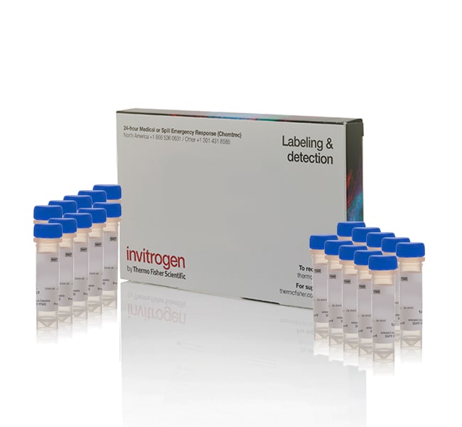
Sondas fluorescentes CellTracker™
Invitrogen™

Sondas fluorescentes CellTracker™

Es un colorante fluorescente adecuado para supervisar el movimiento de las células o su ubicación.
Have Questions?
Cambiar vistabuttonViewtableView
Número de catálogoCantidadTipo de colorante
C702520 x 50 μgCellTracker Green CMFDA
C29251mgCellTracker Green CMFDA
C29271 mgCellTracker Orange CMTMR
C3455220 x 50 μgCellTracker™ Red CMTPX
Número de catálogo C7025
Precio (USD)
-
Cantidad:
20 x 50 μg
Tipo de colorante:
CellTracker Green CMFDA
Ask our AI about this Product
Cell movement and location studies require specialized probes that are nontoxic to living cells and well retained, allowing for multigenerational tracking. The CellTracker fluorescent probes are available in a range of fluorescent colors to match instrument lasers and filters, and to accommodate co-staining with antibodies or other cell analysis probes. These dyes are excellent tools for monitoring cell movement, location, proliferation, migration, chemotaxis, and invasion.

  • Este colorante se conserva bien, lo que permite el seguimiento multigeneracional de movimientos celulares.
  • Los espectros de excitación/emisión verde (492/517 nm maxima) son ideales para el multiplexing con proteínas y colorantes rojo fluorescente.
  • Fácil de usar: retire el medio, añada el colorante, incube 30 minutos y obtenga la imagen de las células.
  • Retención de la señal fluorescente superior a >72 horas (normalmente de tres a seis generaciones).
  • Baja citotoxicidad: no afecta a la viabilidad ni a la proliferación
  • El tinte fluorescente de CMAC CellTracker Blue se ha diseñado para atravesar libremente las membranas celulares hasta las células donde se transforma en productos de reacción impermeabilizantes de membranas celulares.
  • El colorante se transfiere a las células hijas, pero no a las celdas adyacentes de una población.
  • Estable, no tóxico a las concentraciones de trabajo, bien conservado en las células, y brillante fluorescente a pH fisiológico

Adhesión celular, análisis celular, proliferación celular, marcado y seguimiento celular, viabilidad celular y citotoxicidad, viabilidad celular, proliferación y función, Imágenes celulares, ensayos de toxicología celular, quimiotaxis y migración celular, descubrimiento y desarrollo de fármacos, marcado celular general, detección de glutatión, detección de alto contenido (HCS), inmunofluorescencia (IF), tinción y detección de inmunofluorescencia, señalización y homeostomía, señalización, identificación y señalización Seguimiento microbiano, estrés nitrooxidativo, ensayos ADME/Tox basados en dianas, detección de pH

Para uso exclusivo en investigación. No apto para uso en procedimientos diagnósticos.
Especificaciones
ColorVerde
DescripciónCellTracker™ Green CMFDA Dye, 20 x 50 μg
Método de detecciónFluroescent probes
Tipo de coloranteCellTracker Green CMFDA
Emisión517 nm
Intervalo de longitud de onda de excitación492⁄517
Para utilizar con (aplicación)Fluroescence microscopy, cells in suspension, adherent cells
FormularioDry Powder
Peso molecular464.86
Línea de productosCellTracker™
Cantidad20 x 50 μg
Tipo de reactivoCompuestos de seguimiento celular, reactivos de etiquetado celular
Condiciones de envíoTemperatura ambiente
Tipo de etiquetaOtras etiquetas o colorantes
Tipo de productoTinte
SubCellular LocalizationCytoplasm
Unit SizeEach
Contenido y almacenamiento
Almacenar en congelador entre -5 °C y -30 °C
PG2624-PJT8477-COL022341-Media-Promo-Pod-Graphic-Global-F-278x123
four-plex-image-COS7-live-cell zoomed
Have questions about this product? Ask our AI assisted search.
This is an AI-powered search and may not always get things right. You can help us make it better with a thumbs up or down on individual answers or by selecting the “Give feedback" button. Your search history and customer login information may be retained by Thermo Fisher and processed in accordance with our Privacy Notice.

Figuras

Espectros de fluorescencia

Fluorescence spectra

Customers who viewed this item also viewed


Documentos y descargas

Certificados

N.º de loteCertificate TypeDateCatalog Number(s)
3154213Certificate of Analysis13 may 2025C2925
3148319Certificate of Analysis04 may 2025C7025
3123542Certificate of Analysis17 abr 2025C2927
3112785Certificate of Analysis20 dic 2024C7025
2994050Certificate of Analysis04 sept 2024C7025
Se muestran 5 resultados, busque arriba un certificado específico

Hojas de datos de seguridad

Preguntas frecuentes

Calcein, AM and FDA (fluorescein diaceate) are examples of some dyes used for this application. Since these dyes are not incorporated or covalently attached to any cellular components, they may have a short retention time as some cell types may actively efflux the dye out of the cells. The CellTracker and CellTrace dyes include either a mild thiol-reactive chloromethyl group or amine-reactive succinnimidyl ester group to allow for covalent binding to cellular components, providing for better retention. As with any reagent, one should empirically determine retention times for the cell type used.

Find additional tips, troubleshooting help, and resources within our Cell Analysis Support Center.

Calcein, AM is a good choice for cell tracking and as a general cytoplasmic stain. However, it doesn't bind to anything and may be actively pumped out of the cells within a couple hours, which is likely what happened. The retention of Calcein within live cells is dependent upon the inherent properties of the cell type and culture conditions.

For long-term imaging, you may wish to consider a reactive cytoplasmic stains such as CFDA, SE or the CellTracker and CellTrace dyes.

Find additional tips, troubleshooting help, and resources within our Cell Analysis Support Center.

Yes, the CellTracker dyes react with any accessible thiol part of the protein and can be fixed. However, some CellTracker dyes may be attached to small metabolites that can leak from the cell following permeabilization. This can result in decreased fluorescence.

Find additional tips, troubleshooting help, and resources within our Cell Tracing and Tracking Support Center.

The shelf life for CellTracker Green CMFDA Dye is one year after receiving it, when stored desiccated at -20 degrees C.

One possibility is that there is spectral bleedthrough between the dyes. Be sure to check the single-color samples by imaging the red cells in green and imaging the green cells in red, using the optimal imaging settings for the other color. If you see bleedthrough with these controls, then you will have to reduce the dye label concentration to reduce the brightness of the dyes, or choose dyes that are farther apart spectrally. If the issue isn’t bleedthrough, another possibility is that the cells were not adequately washed after staining, allowing some unincorporated dye to remain and label the other cells after they were introduced. Extending washes and wash times should help with this.

Find additional tips, troubleshooting help, and resources within our Cell Analysis Support Center.

Citations & References (341)

Citations & References
Abstract
In situ image analysis of interactions between normal human keratinocytes and fibroblasts cultured in three-dimensional fibrin gels.
Authors:Sun T,Haycock J,Macneil S
Journal:Biomaterials
PubMed ID:16510181
Evaluation of a flow cytometric fluorescence quenching assay of phagocytosis of sensitized sheep erythrocytes by polymorphonuclear leukocytes.
Authors:Van Amersfoort ES, Van Strijp JA
Journal:Cytometry
PubMed ID:7875036
A number of reports have been published describing phagocytosis assays for flow cytometric analysis. In some of these, the fluorescence quenching technique has been used to discriminate between adherent and ingested particles. In this report, we have evaluated the efficacy of a quantitative fluorescence quenching technique with crystal violet and ... More
A point mutation in the binding subunit of a retroviral envelope protein arrests virus entry at hemifusion.
Authors:Zavorotinskaya T, Qian Z, Franks J, Albritton LM
Journal:J Virol
PubMed ID:14671127
The transmembrane subunits of viral envelope proteins are thought to perform all of the functions required for membrane fusion during entry of enveloped viruses. However, changes in a conserved SPHQ motif near the N terminus of the receptor binding subunit of a murine leukemia virus (MLV) envelope protein block infection ... More
Direct priming of antiviral CD8+ T cells in the peripheral interfollicular region of lymph nodes.
Authors:Hickman HD, Takeda K, Skon CN, Murray FR, Hensley SE, Loomis J, Barber GN, Bennink JR, Yewdell JW,
Journal:Nat Immunol
PubMed ID:18193049
'It is uncertain how antiviral lymphocytes are activated in draining lymph nodes, the site where adaptive immune responses are initiated. Here, using intravital microscopy we show that after infection of mice with vaccinia virus (a large DNA virus) or vesicular stomatitis virus (a small RNA virus), virions drained to the ... More
Single-molecule analysis of cadherin-mediated cell-cell adhesion.
Authors:Panorchan P, Thompson MS, Davis KJ, Tseng Y, Konstantopoulos K, Wirtz D
Journal:J Cell Sci
PubMed ID:16371651
'Cadherins are ubiquitous cell surface molecules that are expressed in virtually all solid tissues and localize at sites of cell-cell contact. Cadherins form a large and diverse family of adhesion molecules, which play a crucial role in a multitude of cellular processes, including cell-cell adhesion, motility, and cell sorting in ... More
341 total citations

Compartir número de catálogo, nombre o enlace.

1x1 image pixel for data collection