Search Thermo Fisher Scientific
ReadyProbes Ready-to-Use Imaging Reagents

The easiest way to image your cells
Molecular Probes ReadyProbes reagents are ready-to-use imaging solutions designed to allow you to stain cells with:
- No calculations
- No dilutions
- No pipetting
Reach for the convenient dropper bottle, add 2 drops per milliliter of your cell suspension, and you're ready to image. ReadyProbes reagents address your most common imaging needs, including nuclear and cytoskeletal staining, and analyses for cell viability and apoptosis. In addition, we’ve formulated some of our most popular secondary antibodies for use straight from the dropper bottle. We’ve also developed ReadyProbes imaging accessories to help you get great results from your staining protocol.
On this page:
ReadyProbes Cell Stains
With each ReadyProbes reagent, you can follow the icon-based protocol to obtain quick and easy results for a common imaging application. ReadyProbes cell stains can remain on the bench within arm’s reach of your microscope. Add 2 drops per milliliter of solution, and you're ready for the next step in your experiment.

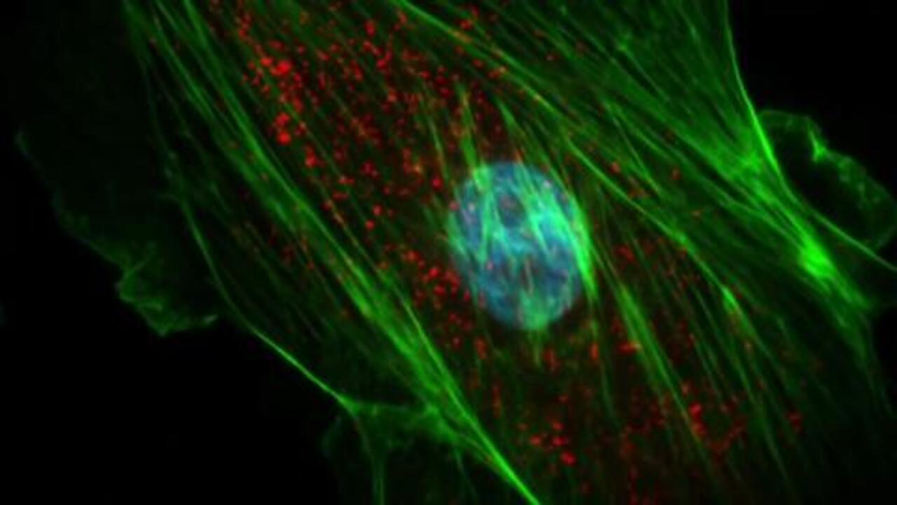
HeLa cells labeled with NucBlue Live ReadyProbes Reagent and ActinRed 555 ReadyProbes Reagent
NucBlue Live ReadyProbes Reagent
|
NucRed Live 647 ReadyProbes Reagent
|
NucBlue Fixed Cell ReadyProbes Reagent
| |
|---|---|---|---|
| Readout |
The easiest fluorescent staining method for nucleic acids; no calculations, no dilutions, no pipetting
| ||
| Target |
High-efficiency staining of both RNA and DNA
| ||
| Common filter set | DAPI | Cy5 | DAPI |
| Ex/Em (nm) | 358/461 | 638/686 | 360/460 |
| Signal-to-noise ratio |  |  | |
| Photostability | | | |
| Live cells |
Yes
|
Yes
|
No
|
| Fixed cells |
Yes
|
Yes
|
Yes
|
| Fixable |
Yes
|
Yes
|
NA
|
| Format |
6 x dropper bottles
|
6 x dropper bottles
|
6 x dropper bottles
|
| Cat. No. | R37605 | R37106 | R37606 |
See more nuclear staining reagents
ActinGreen 488 ReadyProbes Reagent
|
ActinRed 555 ReadyProbes Reagent
| |
|---|---|---|
| Readout |
The easiest fluorescent staining method for actin; no calculations, no dilutions, no pipetting
| |
| Target |
High-affinity staining of F-actin
| |
| Common filter set | FITC | TRITC |
| Ex/Em (nm) | 495/518 | 555/565 |
| Signal-to-noise ratio | | |
| Photostability | | |
| Live cells |
No
|
No
|
| Fixed cells |
Yes
|
Yes
|
| Fixable |
NA
|
NA
|
| Platforms |
Imaging
|
Imaging
|
| Format |
6 x dropper bottles
|
6 x dropper bottles
|
| Cat. No. | R37110 | R37112 |
See more actin staining reagents
NucGreen Dead 488 ReadyProbes Reagent
|
NucRed Dead 647 ReadyProbes Reagent
|
ReadyProbes Cell Viability Imaging Kit (Blue/Green)
|
ReadyProbes Cell Viability Imaging Kit (Blue/Red)
| |
|---|---|---|---|---|
| Readout |
The easiest fluorescent staining method for viability; no calculations, no dilutions, no pipetting
| |||
| Target |
Single-color assays detect dead cells
|
Two-color assays detect dead cells and total cells
| ||
Dead cells stain green
|
Dead cells stain red
|
Dead cells stain green, live cells appear blue
|
Dead cells stain red, total cells appear blue
| |
| Fluorescent label |
DNA stain
|
DNA stain
|
DNA stain
|
Propidium iodide
|
Hoechst 33342
|
Hoechst 33342
| |||
| Standard filter set | FITC | Cy®5 | FITC | TRITC/RFP |
DAPI | DAPI | |||
| Ex/Em (nm) | 504/523 | 642/661 | 504/523 | 535/617 |
360/460 | 360/460 | |||
| Platforms |
Imaging
|
Imaging
|
Imaging
|
Imaging
|
| Format |
6 x dropper bottles
|
6 x dropper bottles
|
6 x dropper bottles
|
6 x dropper bottles
|
| Cat. No. | R37109 | R37113 | R37609 | R37610 |
See more cell viability stains
CellEvent Caspase-3/7 Green ReadyProbes Reagent
| |
|---|---|
| Readout |
The easiest fluorescent staining method for apoptosis; no calculations, no dilutions, no pipetting
|
| Target |
Fluorescence intensity increases with caspase 3/7 activity
|
| Common filter set | FITC |
| Ex/Em (nm) | 502/530 |
| Live cells |
Yes
|
| Fixed cells |
No
|
| Fixable |
Yes
|
| Platforms |
Imaging
|
| Format |
1 x dropper bottle
|
| Cat. No. | R37111 |
See more apoptosis stains
NucBlue Live ReadyProbes Reagent
|
NucRed Live 647 ReadyProbes Reagent
|
NucBlue Fixed Cell ReadyProbes Reagent
| |
|---|---|---|---|
| Readout |
The easiest fluorescent staining method for nucleic acids; no calculations, no dilutions, no pipetting
| ||
| Target |
High-efficiency staining of both RNA and DNA
| ||
| Common filter set | DAPI | Cy5 | DAPI |
| Ex/Em (nm) | 358/461 | 638/686 | 360/460 |
| Signal-to-noise ratio | | | |
| Photostability | | | |
| Live cells |
Yes
|
Yes
|
No
|
| Fixed cells |
Yes
|
Yes
|
Yes
|
| Fixable |
Yes
|
Yes
|
NA
|
| Format |
6 x dropper bottles
|
6 x dropper bottles
|
6 x dropper bottles
|
| Cat. No. | R37605 | R37106 | R37606 |
See more nuclear staining reagents
ActinGreen 488 ReadyProbes Reagent
|
ActinRed 555 ReadyProbes Reagent
| |
|---|---|---|
| Readout |
The easiest fluorescent staining method for actin; no calculations, no dilutions, no pipetting
| |
| Target |
High-affinity staining of F-actin
| |
| Common filter set | FITC | TRITC |
| Ex/Em (nm) | 495/518 | 555/565 |
| Signal-to-noise ratio | | |
| Photostability | | |
| Live cells |
No
|
No
|
| Fixed cells |
Yes
|
Yes
|
| Fixable |
NA
|
NA
|
| Platforms |
Imaging
|
Imaging
|
| Format |
6 x dropper bottles
|
6 x dropper bottles
|
| Cat. No. | R37110 | R37112 |
See more actin staining reagents
NucGreen Dead 488 ReadyProbes Reagent
|
NucRed Dead 647 ReadyProbes Reagent
|
ReadyProbes Cell Viability Imaging Kit (Blue/Green)
|
ReadyProbes Cell Viability Imaging Kit (Blue/Red)
| |
|---|---|---|---|---|
| Readout |
The easiest fluorescent staining method for viability; no calculations, no dilutions, no pipetting
| |||
| Target |
Single-color assays detect dead cells
|
Two-color assays detect dead cells and total cells
| ||
Dead cells stain green
|
Dead cells stain red
|
Dead cells stain green, live cells appear blue
|
Dead cells stain red, total cells appear blue
| |
| Fluorescent label |
DNA stain
|
DNA stain
|
DNA stain
|
Propidium iodide
|
Hoechst 33342
|
Hoechst 33342
| |||
| Standard filter set | FITC | Cy®5 | FITC | TRITC/RFP |
DAPI | DAPI | |||
| Ex/Em (nm) | 504/523 | 642/661 | 504/523 | 535/617 |
360/460 | 360/460 | |||
| Platforms |
Imaging
|
Imaging
|
Imaging
|
Imaging
|
| Format |
6 x dropper bottles
|
6 x dropper bottles
|
6 x dropper bottles
|
6 x dropper bottles
|
| Cat. No. | R37109 | R37113 | R37609 | R37610 |
See more cell viability stains
CellEvent Caspase-3/7 Green ReadyProbes Reagent
| |
|---|---|
| Readout |
The easiest fluorescent staining method for apoptosis; no calculations, no dilutions, no pipetting
|
| Target |
Fluorescence intensity increases with caspase 3/7 activity
|
| Common filter set | FITC |
| Ex/Em (nm) | 502/530 |
| Live cells |
Yes
|
| Fixed cells |
No
|
| Fixable |
Yes
|
| Platforms |
Imaging
|
| Format |
1 x dropper bottle
|
| Cat. No. | R37111 |
See more apoptosis stains
ReadyProbes Secondary Antibodies
ReadyProbes secondary antibodies are conjugated with bright and photostable Alexa Fluor 488 and Alexa Fluor 594 dyes to offer convenient and robust labeling. These Alexa Fluor secondary antibodies recognize IgG heavy chains and all classes of immunoglobulin light chains from either mouse or rabbit. Like other ReadyProbes reagents, these secondary antibodies are provided in a convenient dropper bottle format. No pipetting or dilutions are required. Just add 2 drops per milliliter of buffer to stain your cells, and follow the icon-based protocol.

CAKI cell peroxisomes labeled using a rabbit anti-PMP70 antibody, followed by detection with Alexa Fluor 488 Goat Anti–Rabbit IgG Antibody ReadyProbes Reagent.
See moresecondary antibodies
ReadyProbes sample preparation accessories
Imaging accessories makes it easier to handle cell or tissue samples and to prepare for staining and imaging workflow.

HeLa cells fixed and permeabilized using the Image-iT Fixation/Permeabilization Kit.
Image-iT fixation and permeabilization Kits
Image-iT fixative and permeabilization solutions and kits are ready-to-go solutions for fluorescence imaging. These reagents are manufactured from high quality ingredients, which helps minimize autofluorescence artifacts. These reagents are also free from impurities, such as methanol, which can interfere with cell/tissue morphology. No more need to handle toxic powders or hard-to-mix reagents, these reagents are designed for all fluorophores, including as Alexa Fluor dyes, FITC, TRITC, GFP and mCherry.
General blockers
General blockers are used in workflows such as, immunocytochemistry (ICC), immunohistochemistry (IHC), or in-situ hybridization (ISH) to help reduce off-target binding of antibodies and other probes/indicators. ReadyProbes blockers are generated with high quality ingredients to help minimize autofluorescence artifacts caused by substandard ingredients. Use these reagents after fixative step but before primary or secondary antibody incubations.
Specific blockers
In fluorescence imaging, reagent and specimen interactions can cause background fluorescence artifacts and non-specific signals. To address these issues, we’ve developed specialized blockers to help minimize these artifacts, so the specific signal can be observed.
| Staining/background issue | Probable cause | Solution | Cat. No. |
|---|---|---|---|
| Tissues have background, non-specific fluorescence before adding any fluorophore conjugated reagent to the cells/tissue. | Many tissue types, and some cells have endogenous autofluorescence | ReadyProbes Tissue Autofluorescence Quenching Kit | R37630 |
| Nonspecific signal when adding fluorophore conjugated secondary antibody (without primary antibody) | Static charge on fluorophore-conjugated secondary antibody is causing non-target staining | Image-iT FX Signal Enhancer ReadyProbes Reagent | R37107 |
| Non-specific signal when mouse-originating antibodies are used on mouse tissue | Mouse-originating antibodies target non-specific targets in mouse tissue | ReadyProbes Mouse-on-Mouse IgG Blocking Solution (30X) | R37621 |
| Adding reaction buffer, without HRP/AP–conjugated antibody generates non-specific signal | Many tissue/cells have endogenous peroxidase and/or phosphatase activity | ReadyProbes Endogenous HRP and AP Blocking Solution (1X) | R37629 |
| Adding fluorophore conjugated avidin and/or biotin generates non-specific signal | Many tissue/cells have endogenous avidin and/or biotin, or similar molecules. | ReadyProbes Avidin/Biotin Blocking Solution (1X) | R37627 |
| Adding fluorophore conjugated streptavidin and/or biotin generates non-specific signal | Many tissue/cells have endogenous avidin and/or biotin, or similar molecules. | ReadyProbes Streptavidin/Biotin Blocking Solution (1X) | R37628 |
See morelive-cell imaging accessories
See morefixed-cell imaging accessories
Resources
More information | Related products |
For Research Use Only. Not for use in diagnostic procedures.